Field of Science

No shit - a new way to study diarrhoeal disease

Diarrhoeal disease is awful. I don't think I have to tell anyone that. 

According to the WHO:

Key facts
  • Diarrhoeal disease is the second leading cause of death in children under five years old. It is both preventable and treatable.
  • Diarrhoeal disease kills 1.5 million children every year.
  • Globally, there are about two billion cases of diarrhoeal disease every year.
  • Diarrhoeal disease mainly affects children under two years old.
  • Diarrhoea is a leading cause of malnutrition in children under five years old.

Behold the wonder of the intestine
 The major causes of disease are infectious agents, namely the likes of noroviruses or rotaviruses. (very good wikipedia article here) But also bacteria, parasites and non microbial assaults. These pathogens enter our bodies via the oro-faecal route where they are able to infect the lining of our intestinal tract. Here they exert their biological effects and manipulate their resident tissue to aid in their replication and spread in the general population. In areas with poor sanitation this is why these diseases are such massive killers. They are acutely adapted to this way of life.

We are somewhat out of our grasp when dealing with - and studying - these organisms. In many cases (noroviruses especially) we cannot even grow the viruses in the lab. Many groups use mouse noroviruses and immunocompromised mice but of course this really isn't optimal for human viruses - especially when pathogenesis and drug development/vaccine studies come along.

So, if we cannot grow them how can we study them? Plus even if we did have the ability to culture them we lack accurate model systems of the human gut to even discover anything worthwhile about the way they grow.

But imagine if we had the ability to study human viruses in the lab, which had been themselves grown in the lab, with human tissue which had also been grown in the lab. 


Enter the human intestinal organoid.  

An intestinal organoid a la Spence et al 2011


An organoid is a structure that resembles an organ. It is not an 'organ' itself taken from a body - it's constructed in the lab to function as one. But organs are pretty complex and none less so than the gut. Its complexity resides in the physical (three dimensional) and biological (cell type/gene expression) planes. So how can we build this 'awesome' work of evolutionary engineering ourselves?

Easy.

You just copy mother nature.

Briefly, the NIH-approved embryonic stem cell line WA09 (originating from the WiCell Research Institute and obtained from the Baylor College of Medicine Human Embryonic Stem Cell Core) was cultured using feeder-free conditions. Stem cells were split at a high density, and, once they reached 85 to 90% confluence, cells were treated for 3 days with a series of differentiation media containing activin A to begin differentiation into definitive endoderm. Definitive endoderm was then treated for 2 to 5 days with growth factors Wnt3a and FGF4, leading to formation of hindgut spheroids. Once spheroids spontaneously detached from monolayers, they were collected, embedded into matrigel (BD Biosciences), and supplied with media supplemented with intestinal growth factors (Wnt3a, R-Spondin1, Noggin, and epidermal growth factor [EGF]; all supplied from R&D Systems). Spheroids matured into intestinal organoids over the course of ~1 to 2 months before they were used for experiments.
It started off like this


Anded up like this
Grown in this way you can 'easily' generate what effectively looks and feels like a human intestinal epithelium (these things also contain some underlying mesenhcymal tissue). They can even be kept alive for over 3 months and the stem cells from which they derive can be frozen and re-animated any time to set up more and more organoid cultures.  


So do these organoids allow viral replication? And can we use them to understand how these viruses infect and cause disease? Well the short answer is yes.


A U.S group from Baylor College of Medicine in Texas were able to cut open the spherical structures and infect them with rotaviruses, even clinical isolates of the virus (OA paper here). In this case the virus needed access to the inside of the structure, the part the resembles in insides of our gut and the place where the cell it likes to replicate in are found. These viruses bound to cells, got in and began replicating and generating new viral particles which could go on and initiate a whole new round of growth. This a whole lot better than using primary monkey kidney cells to isolate and grow the virus or using lab adapted strains.


Organoid structures could be infected with rotaviruses


This was only a preliminary observational study showing a proof of concept that this technique which had previously only been done using mouse stem cells, could work for humans and that they could be used for infection studies. What they didn't show was that rotavirus infection of the organoids bore any resemblance pathogenically to infection in humans. I'm sure this is the next step. What also would be of great use would be to see whether other non-culturable but important human pathogens could be grown this way - I'm thinking noroviruses. What is neat about this work is that we have the ability through recombinant gene vectors to knockdown or over-express any gene we want. The initial paper documented this in 2011. And this is being done with an NIH-approved stem cell line - imagine what could be done with healthy or 'diseased' stem cells.

One problem with this lies in the way they were infected. In order to access the inside of the spherical structure the organoid was cut open using a tungsten needle - an extremely sharp instrument. Who know what kind of effect this would have on nearby cells? But I guess without it this work could not be done and this was probably the safest way of doing so.


Another issue is - like the majority of other in vitro models - there is not immune system component to the structure. And of course the innate immune cells residing in tissues would have a wide ranging effect on the subsequent spread and infection kinetics of the virus. However, you could imagine that in the future it may not be too difficult to add these in to the system. 


ResearchBlogging.orgStacy R. Finkbeinera,, Xi-Lei Zenga,, Budi Utamaa,, Robert L. Atmara,b,, Noah F. Shroyerc,, & and Mary K. Estesa,b (2012). Stem Cell-Derived Human Intestinal Organoids as an Infection Model for Rotaviruses mBio, 3 (4) DOI: 10.1128/mBio.00159-12

#microtwjc 5(?) Microbiology of the built environment - the toilet edition


This weeks microbiology twitter journal club is:


Microbial Biogeography of Public Restroom Surfaces


Gilberto E. Flores1, Scott T. Bates1, Dan Knights2, Christian L. Lauber1, Jesse Stombaugh3, Rob Knight3,4, Noah Fierer1,5*
1 Cooperative Institute for Research in Environmental Science, University of Colorado, Boulder, Colorado, United States of America, 2 Department of Computer Science, University of Colorado, Boulder, Colorado, United States of America, 3Department of Chemistry and Biochemistry, University of Colorado, Boulder, Colorado, United States of America, 4 Howard Hughes Medical Institute, University of Colorado, Boulder, Colorado, United States of America, 5 Department of Ecology and Evolutionary Biology, University of Colorado, Boulder, Colorado, United States of America

have a look at the abstract here (emphasis my own):
We spend the majority of our lives indoors where we are constantly exposed to bacteria residing on surfaces. However, the diversity of these surface-associated communities is largely unknown. We explored the biogeographical patterns exhibited by bacteria across ten surfaces within each of twelve public restrooms. Using high-throughput barcoded pyrosequencing of the 16 S rRNA gene, we identified 19 bacterial phyla across all surfaces. Most sequences belonged to four phyla: Actinobacteria,BacteriodetesFirmicutes and Proteobacteria. The communities clustered into three general categories: those found on surfaces associated with toilets, those on the restroom floor, and those found on surfaces routinely touched with hands. On toilet surfaces, gut-associated taxa were more prevalent, suggesting fecal contamination of these surfaces. Floor surfaces were the most diverse of all communities and contained several taxa commonly found in soils. Skin-associated bacteria, especially the Propionibacteriaceae, dominated surfaces routinely touched with our hands. Certain taxa were more common in female than in male restrooms as vagina-associated Lactobacillaceae were widely distributed in female restrooms, likely from urine contamination. Use of the SourceTracker algorithm confirmed many of our taxonomic observations as human skin was the primary source of bacteria on restroom surfaces. Overall, these results demonstrate that restroom surfaces host relatively diverse microbial communities dominated by human-associated bacteria with clear linkages between communities on or in different body sites and those communities found on restroom surfaces. More generally, this work is relevant to the public health field as we show that human-associated microbes are commonly found on restroom surfaces suggesting that bacterial pathogens could readily be transmitted between individuals by the touching of surfaces. Furthermore, we demonstrate that we can use high-throughput analyses of bacterial communities to determine sources of bacteria on indoor surfaces, an approach which could be used to track pathogen transmission and test the efficacy of hygiene practices

Here's my thoughts of this (briefly) 

As you can see from this paper certain American restrooms harbour a massive diversity of bacterial species (only bacteria were looked at - I wonder what kind of viruses are here). The majority of which have a strong association with the human species; others come from soil or water. Most of all the diversity of bacteria in the restrooms come from our skin (no surprise there really). 

You might ask: "So what?", "Who cares?" or "Why bother?" 

Is it just because you had access to some fancy machine and 12 bathrooms?

I have to disagree with this viewpoint: This is only the beginning.

 In order to fully understand the microbiology of the built environment and how it effects the human population residing in it we have to start somewhere - and deep sequencing or bacterial diversity is a damn good place t start. (after all we spend the majority of our time in doors - and even when we're outside it will probably be in or in close proximity to major population centres like cities). 

If you don't believe have a look at this site.

This falls into a larger investigation looking into the microbial diversity found across different environments, such as the office (this groups follow on paper) or nursery. We can study 'healthy' environments and we can study 'unhealthy' environments. Potentially there are important microbiological differences between the two. By determining the baseline microbiome we could in theory begin to specifically alter its appearance to enhance the human experience of the build environment for health, environmental or economic reasons. 

What's the difference between one kid with a fever and one without?

A paper out last month goes a little way to answering a question I have had for a while: what are the major causes of fevers in children?

Imagine this: your child goes to the doctor, they have a fever so they might get given antibiotics or they might be sent home for bed rest because of a non-descript  'viral' infection. But what is that non-descript virus? and how is it causing feverish disease? Maybe if we knew exactly what was doing this we may have some chance to prevent it.


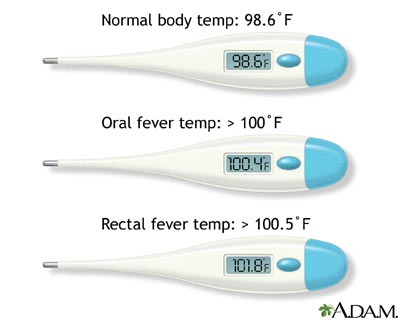
Fevers are an annoyance and in some cases can even be life threatening. Often times they are your bodies way of helping clear an infection. Your body will 'see' and detect infectious (and potentially dangerous) micro-organisms like viruses or bacteria and set off a chain reaction of chemical signals resulting in a rising temperature. But fevers may also be caused by a range of other ailments: arthritis, cancer or basically anything that causes inflammation. 


This paper in PLoS ONE suggests that we should delve into those children's viromes to see what is making them sick. Using next generation sequencing of nasopharyngeal swabs as well as plasma blood samples from febrile and non-febrile American children, they attempt to tease apart they causes of their illness. They also looked with PCR to detect viruses more specifically. This allowed them to essentially cover all viral bases in a comprehensive manner and hopefully detect what was truly in there. 


However, this approach is only based on looking for viral genetic sequences and no search for the actual virus was carried out. This means that those viruses detected could may never have infected the person, could have come directly from the environment or had been recently cleared by the immune system. WIthout virus isolation we cannot be sure to what extent a viral sequence is associated with a certain condition.
The viruses they found by PCR and deep sequencing.

In some cases this knee-jerk reaction to infection actually can damage you and can even result in seizure and death. Diagnosing the cause of fevers and treating early in thus a major concern and of the major issues of fevers is that they can be set off by a multitude of pathogens, the majority of which we have never really studied in much detail. Hence this group spending their time and money investigating it. 


Abstract:

Why did they do the work:

Unexplained fever (UF) is a common problem in children under 3 years old. Although virus infection is suspected to be the cause of most of these fevers, a comprehensive analysis of viruses in samples from children with fever and healthy controls is important for establishing a relationship between viruses and UF.


Methods:


We used unbiased, deep sequencing to analyze 176 nasopharyngeal swabs (NP) and plasma samples from children with UF and afebrile controls, generating an average of 4.6 million sequences per sample. 


Comparing viruses seen in febrile versus afebrile kids. Spot the differences?
Results:


An analysis pipeline was developed to detect viral sequences, which resulted in the identification of sequences from 25 viral genera. These genera included expected pathogens, such as adenoviruses, enteroviruses, and roseoloviruses, plus viruses with unknown pathogenicity. Viruses that were unexpected in NP and plasma samples, such as the astrovirus MLB-2, were also detected. Sequencing allowed identification of virus subtype for some viruses, including roseoloviruses. Highly sensitive PCR assays detected low levels of viruses that were not detected in approximately 5 million sequences, but greater sequencing depth improved sensitivity. On average NP and plasma samples from febrile children contained 1.5- to 5-fold more viral sequences, respectively, than samples from afebrile children. Samples from febrile children contained a broader range of viral genera and contained multiple viral genera more frequently than samples from children without fever. Differences between febrile and afebrile groups were most striking in the plasma samples, where detection of viral sequence may be associated with a disseminated infection. 


Conclusions:


These data indicate that virus infection is associated with UF. Further studies are important in order to establish the range of viral pathogens associated with fever and to understand of the role of viral infection in fever. Ultimately these studies may improve the medical treatment of children with UF by helping avoid antibiotic therapy for children with viral infections.

Points to consider:

As I said previously, the group only looked at viral sequences. No viruses were isolated or grown from these samples which makes it a little complicated in drawing conclusions based on this data. But it is extremely interesting and worthwhile and perhaps in some instances may give us a less biased view into the virome.

We all harbour viral sequences. Even the 'healthy' control populations when you look at their upper respiratory tract and circulation, indicating that the issue of fever versus no fever may be more complicated than expected. Maybe some people are much easily able to withstand disease progression from an infection.

They only looked at plasma and at the upper respiratory tract. Many other parts of the body could be sampled in the future to more comprehensively investigate how the virome of a particular is associated with any particular condition or disease. The digestive tract, urogenital tract or blood cells (not plasma) could and should be investigated.

Causation cannot be determined from this study. And they even say that this was not the aim of the investigation. To determine causation that a particular virus caused fever in kids one would have to fulfil Koch's postulates.

Fever could have been associated with bacterial exposure/infection yet this was not looked at here. Perhaps this may have been more obvious to the diagnosing clinician and antibiotics would have prescribed.

In conclusion, this is an interesting paper but it is in a sense quite preliminary. They have defined their methods and proven they can detect a wide range of viral sequences in clinical samples. But at the minute it is rather difficult to conclude anything else from this. I look forward to further characterisation of the viromes of multiple other tissue samples and even the characterisation of the viruses they identified. Perhaps they will represent entirely unknown disease causing viruses.


Kristine M. Wylie1*, Kathie A. Mihindukulasuriya1, Erica Sodergren1, George M. Weinstock1, Gregory A. Storch2 (2012). Sequence Analysis of the Human Virome in Febrile and Afebrile Children PLoS ONE, 7 (6) : 10.1371/journal.pone.0027735

Merck's mumps vaccine is good. But not good enough.


It came out this week that two former employees are suing Merck (here's the document), the pharmaceutical company/giant, over what they say was a deliberate attempt to con the US government - and by extension many other countries - into buying a mumps vaccine in the form of their combination MMR-II vaccine that was not as effective as they had previously stated. 


As documented here this has been jumped on by a number of anti-vaccinationists but if you really look at the data from the "real world", you'll quickly see how all parties have really jumped the gun a bit on this issue and that clearly the vaccine is - and has been - working well. But it could be better.

There's no evidence for NI Health Minister lifetime ban on MSM blood donation

It's not often that I talk about politics on this blog. In fact I don't think I have even mentioned it before but in some instances when infectious diseases and politics collide I feel I have to do so. 


One such instance happened today when Edwin Poots a local politician in the Northern Ireland Assembly finally released his decision on whether our blood donation services would accept blood from men who have sex with men. We had been waiting nearly a year for this. 


Edwin Poots, NI Health Minister


We cannot accept blood from men who have sex with men


It's safe to say that sadly Edwin came to the decision that our local Health Service cannot accept blood from men who have sex with men (MSM). In fact he would have even wished that this whole-life ban on blood donation was extended to all 'high risk groups'. 

"I think that people who engage in high-risk sexual behaviour in general should be excluded from giving blood," the minister said.
"And so someone who has sex with somebody in Africa or sex with prostitutes, I am very reluctant about those people being able to give blood."


This was released in an interview for a national news program. Normally I ignore the ramblings of local politicians but in this case Edwin is our Health Minister and at the end of the day has control over the kinds of health services we get.


His decision, which flies in face of what has happend in England, Scotland and Wales who reduced their ban to 12 months post sex (see below), was based upon two pieces of research that "strengthened his position". He never gave any more details about this work which worries me slightly. Now I like the idea that our politicians are basing decisions upon evidence but I'm not sure this research actually exists and if it does it clearly made little impact on the independent review that came out last year (see below).


Poots' statement has rightly drawn criticism from other politicians who correctly state that we have a robust screen program for all blood coming in and brought up that it is not right to discriminate like this. 


But we really need more blood


All this news come at a rather poor time only a week after UK's National Blood week and World Blood Donor Day. These events are set up to celebrate those who actually give blood products and looks to inspire and encourage more and more people to do so. This is necessary because we are in a pretty bad situation here in the UK and in Northern Ireland especially. In fact we even have to import blood in from England, Scotland and Wales to cover our needs. The stat is that only about 5% of all people who CAN give blood do so. And here is a UK health minister effectively turning down blood from the 1.8 million gay or bisexual men across Northern Ireland. Edwin Poots stated that it's potentially unsafe for these "at-risk" peoples to donate blood but is this really true?


As a little background to this story, in September 2011 the health ministers across England, Scotland and Wales - spurred by independent research (see here)- finally lifted the lifetime ban on blood donation from MSM. 


The data from the report was: 


Risks of transmission*from the BBC report

  • Lifetime exclusion - risk of one infection in every 4.41 million donations
  • Five-year time limit - risk of one infection in every 4.39 million donations
  • One-year time limit - risk of one infection in every 4.38 million donations
  • No limit - risk of one infection in every 3.48 million donations
You can see that 1) NO exclusion criteria are perfect. Even with lifetime exclusion there is still some risk of blood-donation infections. But also 2) that changing from a lifetime to a 5 or 1 year limit barely increases the risk of infection (from one in 4.41 million donations to one in 4.38 million donations). This translates into an increase of one infection per 30,000 donations and was deemed to be insignificant given the number of new blood into the transfusion services.


They changed it so that only those men who have had homosexual sex in the last 12 months would be stopped donating. The previous ban had been implemented from the 1980's before the time of HIV testing. Currently all blood is screened for evidence of HIV, hepatitis B and C infection through the detection of anti-viral antibodies. The problem with this method of testing is that in some people it can take up to a year to develop antibodies against a particular virus. That means that if you were to test someone and they showed up negative you couldnt be entirely certain whether they were or weren't virus infected. This is known as the 'window-period' of HIV infection and hence the 12 month restriction which limits the numbers of false-negative results. 


What about the evidence


As the ability of virus testing got better and better we were able to reduce the indefinite ban on MSM donations which along with equality issues made up the impetus to lift the ban last year. There is even evidence to back this safety record up from an Australian study reported in 2010


No evidence of a significantly increased risk of transfusion-transmitted human immunodeficiency virus infection in Australia subsequent to implementing a 12-month deferral for men who have had sex with men.


Here's more evidence,


Relative risk of reducing the lifetime blood donation deferral for men who have had sex with men versus currently tolerated transfusion risks.


And even more:


Scientific background on the risk engendered by reducing the lifetime blood donation deferral period for men who have sex with men.


The 12 month ban on blood from MSM brings them up to date with the recommendations for blood from other "at-risk" groups. But the problem with this is that even still those MSM are still more discriminated against than a heterosexual male who has had multiple instances of unprotected sex. This position is defended because the increase in risk of infection (see data above) is deemed to significant and unwarranted. Despite this people have called that our blood donation deference criteria should be based on individuals and not on groups.  


But the England, Scotland and Wales 12 month ban is certainly a step in the right direction in terms of providing better care for those people who need blood and in limiting discrimination against MSM. So why is Northern Ireland different? I am really not sure but what I do know is that it will definitely negatively impact our local health services by the reducing the pool of safe blood to be transfused. In light of not seeing our Ministers' evidence I cant really comment on it. Maybe he'll release it. Maybe. What I do hope is that this act has not been motivated by personal religious views and bigotry which has cropped up time and time again.


Stupid spam posts

Apologies to anyone who has had their RSS feeds filled with very annoying (and sometimes arabic) spam posts coming from this blog. Hopefully I have cleared this mess up and if I haven't, I apologise for the future. Thanks to all you guys who noticed this and contacted me. The irony of a spam virus on a virus blog is not lost on me!

The origin of Schmallenberg Virus and the need for more surveillance.

ResearchBlogging.orgLast week we finally got the answer to where Schmallenberg virus came from. At least genetically speaking that is (we still don't know from what geographical region it was nor whether it had been in Europe this whole time). But we do now have some clues.


It has come to light that this previously unheard-of microbe is a mixture of two previously known and closely related viruses: Sathuperi and Shamonda viruses. Writing in Archives of Virology earlier this month, a Japanese group (Yanase et al, from the Japanese National Institute of Animal Health) delved into the depths of bunyavirus genetics and uncovered Schmallenberg's closest cousins by sequencing a number of other viruses from Africa and Australiasia. Sadly this paper is not open access.


I wrote about Schmallenberg virus soon after it was discovered earlier this year. This was the virus that popped up in sheep and cows last summer then by the time the next Spring came we quickly realised it's aftermath: it had induced a number of malformations in their young - who at the time of infection were in the womb. These often times had fatal consequences. 


Back then we had no idea where this virus had come from as it's genome sequenced really looked like nothing we had seen previously. The original paper only used one small part of the viruses genome to trace it's ancestry due to the low amount of sequence data for these viruses. Although it bore a distant relation to known viruses there were some significant gaps in our knowledge of genomes from this group of viruses.


A bunyavirus (from ViralZone). Note the three segments of genome.


You see Schmallenberg is a Bunyavirus, a group of single-stranded, negative-sensed RNA virus. But the special thing about these guys is that they are segmented. Just like influenza is. And we all know what flu likes to do with it's segmented genome: it likes to reassort and swap bits and pieces of it's self around. A bit like virus sex. Well Bunyaviruses do this as well and it turns out so did the direct ancestor to Schmallenberg.


The three segmented genome. Schmallenberg had the S and L of Shamonda and the M or Sathepuri. (From Viralzone)

When the Japanese group compared the Schmallenberg genome to those of the other viruses that they had just sequenced, the true ancestry of this deadly virus emerged. It was strikingly clear that it's entire genome did not share the same genetic history. Two of it's three segments were very closely related to the Shamonda viruses while the last segment seemed to have a different story to tell: it was more closely related to another, distinct virus called Sathepuri virus.


All this indicates is that at some point in time, two different viruses (Shamonda and Sathepuri) infected the same cell - maybe in an insect, maybe in a mammal - out came a entirely, never-before-seen virus. This virus somehow made it's way to North-West Europe and started infecting various biting insects and farm animals. We can't yet say where this occurred, nor can we say when but what we can say is that it definitely happened. We also can't be sure of what genetic changes the virus had to make in order to function as this kind of chimera and for it to spread into a new geographic niche.


The situation from Influenza (From Virology Blog). Just the same 


The one issue with this work is that of undersampling. We know so little about the genomic diversity of this group of virus and currently have very little data to compare Schmallenberg to. What we need is to sequence a whole range of isolates from across all continents in order to truly answer the question of where this virus originated. 


And even then, this information will be of little use and may even be used to point the finger of blame. We need to hope that Schmallenberg doesn't come back again in the next couple of years and then if it does, we are ready for it this time. The only way this will happen is with increased global recognition and surveillance of these viruses. 


Reference:


Yanase, T., Kato, T., Aizawa, M., Shuto, Y., Shirafuji, H., Yamakawa, M., & Tsuda, T. (2012). Genetic reassortment between Sathuperi and Shamonda viruses of the genus Orthobunyavirus in nature: implications for their genetic relationship to Schmallenberg virus Archives of Virology DOI: 10.1007/s00705-012-1341-8

A virologist's take on the black death genome #microtwjc 2

ResearchBlogging.orgThe second paper in the Microbiology Twitter Journal Club (Tuesday the 22nd May 2012) is the paper out last year documenting the sequencing and assembly of the complete genome of a strain of Yersinia Pestis (plague) from a 14th Century burial site. It's open access so check it out here.


Here's a PBS news story (w/ interview with lead author as well):


There's also an accompanying feature covering the history and background to Black Death research by Nature, here. Also includes some nice criticisms of this work too. There's also a Nature Blog article here. And here's New York Times article. There's a range of articles by Michelle Ziegler over at Contagions blog to have a look at. And finally before I forget, Vincent Racaniello's 'This Week in Microbiology' podcast covered it last year here.


Here's the video abstract from Nature:


A third dose of MMR is safe but do we need it?


ResearchBlogging.orgIt was recently reported - at the National Foundation for Infectious Diseases 15th Annual Conference on Vaccine Research - that the rate of adverse effects from a third dose of the measles, mumps and rubella (MMR) vaccine is the same as those of the second dose. This was conducted as part of a Centres for Disease Control study and led by Glen Abedi, an epidemiologist at the CDC and Masters student. I'm basing this on a media report of the conference as the paper has not yet been published. You can find more results here.


This is the first study to look at the safety of receiving a third dose of the vaccine in school children during an outbreak. The study has some obvious caveats but what it brings up is the question of whether we should extend 3-dose coverage to the population as a whole?


A mumps outbreak from 2009 to 2010 in and around New York City offered the CDC the chance to specifically look and see just how safe the administration of the third dose would be. In this outbreak - and in others - a very large percentage of those with clinical mumps had received two doses of MMR. We don't really know why, maybe it's a question of them getting a large dose of mumps, or maybe it's the vaccine not being perfect and inducing waning immunity.


They used this as a booster shot against the mumps virus in order to prevent further infection and spread from the community and to do so, they set up vaccination clinics in a number of school in the Orange County area of NYC. Those immunised were between 11 and 17 years old. These booster clinics have been set up before for mumps. These clinics are very money intensive and have reported to cost around $500/person. 


As a means to stem the tide of the outbreak, this three dose schedule worked and the rates of mumps infection dropped from 4.93/1000 to 0.13/1000. Although there was no reporting of antibody levels before and after the 3 doses. And we can't be sure whether the outbreak would have abruptly ended like this without vaccine intervention. One caveat with these kinds of studies is that they usually administer the dose late into the outbreak. If done earlier they may have completely controlled it's spread.


But to determine whether this was safe they had to send out questionnaires to the families of the kids, they consulted local clinics and they looked up the Vaccine Adverse Events Reporting System (VAERS). Over 90% of those immunised responded and only 115 reported adverse effects 2 weeks after vaccination. Those effects were only local pain/swelling at the injection site, muscle pain and dizziness/light-headedness. All the kinds of things that suggest that your immune system recognised the vaccine. Note that no cases of meningitis, glandular swelling  or orchitis were recognised, somewhat more serious effects of mumps vaccination in some cases.


This research highlights that in certain situations (a relatively small localised outbreak, with very targeted vaccination of schoolchildren) a third dose of MMR is safe. Of course if they wanted to definitively test this I think they would need a bigger sample size/diversity than the 1755 religious school kids. Remember also that they didn't report looking into levels of mumps immunity so time will tell whether these children were really protected and whether they may still get mumps in the next couple of years.


But the question now remains is whether or not we should extend three doses to the general population or even in cases of a localised outbreak. Mumps is a very infectious virus and hence you need very high levels of population protection to achieve herd immunity (estimated as high as 92%). In the U.S, those between 13 and 17 have an estimated 2-dose MMR coverage of 90.5% and the MMR vaccine uptake percentage in colleges etc is just shy of 90%. So maybe what this data says is that in general we should really focus on achieving very high levels of 2-dose MMR but in the cases of a mumps outbreak we could use a targeted third dose. If we didn't mind the cost. Maybe in the future to lower the costs, all schools/campuses will introduce a MMR catch-up when the new students start.


Another issue apparent is whether we need a new mumps vaccine. Clearly our current mumps vaccine has been amazingly effective to date but maybe it is not enough to completely eradicate the virus. This is something we will have to consider now that people are developing newer vaccines against the virus.


Centers for Disease Control and Prevention (CDC) (2010). Update: mumps outbreak - New York and New Jersey, June 2009-January 2010. MMWR. Morbidity and mortality weekly report, 59 (5), 125-9 PMID: 20150887

Viruses This Week in Bats

OK they're cute, but are they deadly?
I was lucky enough to appear alongside the guys over at This Week in Virology (thanks Vincent, Alan, Rich and Dickson) to discuss the recent publication of a paper (here in open access) which identified bats and rodents as potential animal reservoirs for a whole load of newly discovered RNA viruses, among other things. Check out the link above and enjoy what was an extremely fun and interesting hour and a half long experience. 

The paper, from a large group of authors right across the world (actually when you look at where they looked, it wasn't all that much of the globe but I guess it is a sample after all), looked specifically for paramyxoviruses in bats and in rodents. 

They picked paramyxoviruses because these viruses have been known to jump species from mammals into humans and other animals, they cause significant diseases (measles, mumps, respiratory infection and encephalitis and finally because I guess they had to focus somewhere. They actually tried unbiased 'deep sequencing' and quickly found it was heavily biased toward not finding paramyxoviruses. Possibly explaining the lack of paramyxovirus discovery in previous non-targeted efforts.

Their thinking was that these bats and rodents would have the potential to host a large number of viruses due to their high population sizes/densities, close contact with each other and potential to travel large distances (in the case of bats). Of course based on these criteria they could have looked in fish or birds but bats and rodents being mammals, there's a higher likelihood that their viruses could do really well in humans (although look at influenza and human metapneuomovirus). Although I bet you would find hundreds more if you explored the seas and the skies as opposed to the jungles.

They looked in over 10,000 individual animals from 15 places around the world, mostly in the tropics and were able identify 66 previously unknown 'species', more than doubling the potential number of potential paramyxoviruses previously known. They even identified the possibly first cousins of many of our deadly viruses, like mumps, nipah and respiratory syncytial virus. 

They then took this further and looked at how viruses like these grew in bats, whether they caused disease and were the excreted and transmitted within and between bat populations. Instead of exploring what viruses were simply present, this group was more interested in establishing whether bats acted as an animal reservoir as knowing this would be excellent from a public health perspective. For example, what areas/species should be protecting and avoided from human contact.

Also from a purely biological perspective, if we assume that these closely related viruses (for example the 'bat mumps' virus) are well adapted to bats and not to humans and then vice versa for the human viruses, then the similarities and differences in functions of each component of the virus should be illuminating understanding how viruses jump species.