Field of Science

Showing posts with label antivirals. Show all posts

Targeting antivirals to the host, part 2 - success?

Viral pneumonia (from influenza possibly) results from an accumulation of immune cells and fluid in the lung tissue. But how can we stop it?

  In the northern hemisphere it's coming up to 'flu season - the time when many of us will be getting our yearly influenza vaccinations. In the coming months during the thousands of potential viral infections, some form of disease/clinical symptoms may set in (maybe not if you're vaccinated) - think of that headache, cough, runny nose, etc.  We can think of these as a function of both the virus replicating and your response (i.e the host) to that same infection.  And, in infections such as influenza, the ramping up of the cellular immune response in an organ like the lung is responsible for much of the clinical course of the disease (the pneumonia - see above).

  The strange thing is that a lot of our therapies are only targeted to the virus aspect (antivirals) of this: blocking particular enzymes, receptor binding molecules etc. But, what if we could target the host response? And through doing this eliminate disease, while potentially allowing the virus to infect, replicate and ultimately use us without inflicting damage. 

How come direct cell-cell spread of HIV allows ongoing replication in the face of antiviral therapy?

Our struggle with HIV/AIDS epitomizes societies' millennia-old fight with microbial pathogens. One goal of HIV research is to generate effective interventions that will allow us to: 1) prevent further spread of HIV (vaccines and behavioural changes), and 2) eliminate the  virus from those already infected (antivirals).

This sterilizing immunity - as it is referred to - has been a long sought-after goal for a number of viruses, yet widespread use of highly-active anti-retroviral therapy (HAART)  fails to completely remove the virus from an individual patient. Somewhere, somehow HIV is continuously replicating in your body. But how and why is this possible?

The spread of HIV from Dendritic cells to T lymphocytes or T to T cells may allow escape from antivirals. http://pathmicro.med.sc.edu

One potential mechanism is that HIV lies in a latent state - anatomical (central nervous system) or biochemical (following DNA integration and before gene expression) - where these drugs cannot inactivate it.

But researchers, headed by David Baltimore at the California Institute of Technology, have come forward with both theoretical and experimental evidence suggesting a novel mechanism that explains how the virus may be able to circumvent HAART treatment through continuous replication and direct cell-cell spread. Their results were published in Nature last week. See here.

What is cell-cell spread?

Viruses can infect new cells via a number of mechanisms. The most well-characteristic being via cell-free virus particles (see computer simulation paper here). In this, new viruses are released from the originally infected cell and diffuse to an uninfected one nearby, thus establishing a novel infection.The problem with this is that it is pretty inefficient. For one, the viruses could be carried anywhere and even if they reach a cell, it may not be the right one. One other, more efficient means of transmission is through direct cell-cell spread, which generally takes diffusion out of the picture.
Model of how HIV moves from cell-cell. A) Dendritic cell (DC) with natural fold-like projections. B) DC picks up virus particles in red C) virus is held in vesicles within cell, D) T cell projections induced, E) formation of the virological synapse, F) release, binding and entry of HIV into T cells. (Felts, et al 2010)

HIV and its close relative, the human T lymphotropic virus (HTLV-1) have been shown to move from one cell to another through this mechanism.

Original paper here

Initial visualisation

In-depth 3D structural analysis

Check out the great videos here


Following interaction with dendritic cells or T cells, the virus directs the assembly of a structure (referred to as the virological synapse) linking the two cells via alterations of the cytoskeleton which causes the two plasma membranes to come close together. This structure initially derives from the target cell.

It is here where new virus particles are released into the gap that has formed, thereby increasing the efficiency of spread by directing transmission and limiting the effects of diffusion.This synaptic structure, ultimately acts to concentrate both virus and receptors at defined subcellular locations. This kind of spread may allow the virus to become essentially invisible to our bodies' immune defences, such as neutralizing antibodies, and even antiviral drugs.

How does this allow escape from treatment?

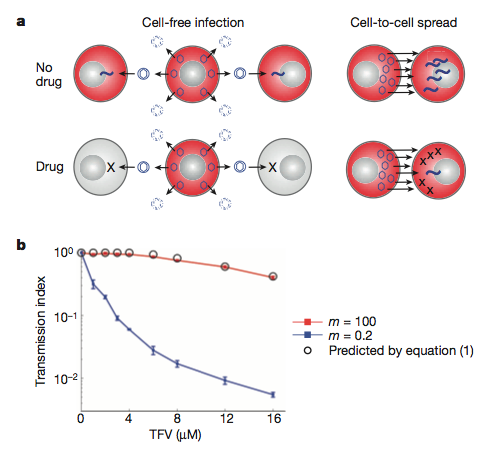
Their model: a) inefficient cell-free infection will be eradicated with antvirals. The more efficient direct cell-cell spread will persist as it involves many more virus particles. b) The mathematical model (backed up by experimental data) showing the loss of infection (transmission index) with increasing concentration of antiviral (TFV) when we have few (m =0.2) or many (m = 100) virus particles.
Expanding the model

Initially, they generated - and experimentally verified - a mathematical model of how HIV transmission may differ during antiretroviral treatment under cell-free (inefficient - low virus transmission) or direct cell-cell spread (efficient - high virus transmission) mechanisms. Their results indicate that when there are many viruses around, the infection is more resistant to the drugs. This, they suggest, is due to the increased probability that at least one virus particle will not interact with the antiviral.


This observation extended to direct cell-cell spread when they introduced previously infected cells with non-infected ones and measured transmission of virus. Both the experimental and theoretical modelling suggest that this spread could be responsible for the ongoing replication seen in patients being treated with antivirals.


What does this mean for HIV therapy? Well, this work brings experimental evidence on the already known process of ongoing HIV replication in the face of antivirals. Through experimentally identifying this mechanism, the researchers here have uncovered a weak-point in HIV biology, one that is not currently being targeted. This method of cell-cell spread may also facilitate escape from neutralizing antibody responses through physically preventing their interaction with HIV proteins. How then might our own bodies combat this virus if its ability to more from cell-cell was inhibited? This work further adds to the growing body of work highlighting direct cell-cell spread as a principle mode of transmission for these retroviruses.

ResearchBlogging.orgSigal A, Kim JT, Balazs AB, Dekel E, Mayo A, Milo R, & Baltimore D (2011). Cell-to-cell spread of HIV permits ongoing replication despite antiretroviral therapy. Nature PMID: 21849975

When you say flu, what do you really mean?

Everyone knows the 'flu when they see it. Its like the cold but only worse, right? But often the public are very quick to brand something the flu when it clearly isn't and this even extends to doctors in the clinic. As highlighted by this recent paper, published in BMC Infectious Diseases.

BioMed Central | Full text | During the summer 2009 outbreak of "swine flu" in Scotland what respiratory pathogens were diagnosed as H1N1/2009?

Turns out a lot of viral infections look like influenza, clinically. But what are doctors misdiagnosing as flu? Based on this study, which paired molecular diagnostics with what the clinic thought each patient had  based on well-defined guidelines (mostly flu at the early stages of the 2009 swine-influenza pandemic - April - July).


Are these symptoms clinically specific to influenza infections? http://symptomswineflu.com 


What they did was:


We examined the results from 3247 samples which were sent to the laboratory during April-July 2009
And,
Real-time RT-PCR was carried out in order to detect influenza A (a generic assay and a H1N1/2009 specific assay [7]), B and C, RSV, rhinovirus, parainfluenza 1-4, human metapneumovirus, coronavirus (229E, NL63, HKU1 and OC43), adenovirus, and Mycoplasma pneumoniae.
 Out of the 3247 samples analysed, 27.9% were diagnosed as being infected with a respiratory pathogen they looked for, what is causing their symptoms??. While human rhinovirus infections were the most common, H1N1 influenza infection was one of the least common at 0.8%. A whole heft of viral and bacterial pathogens were detected at levels intermediate to these.


So, whats going on here? How come it's so difficult to diagnose these guys and after-all, does it really matter?


Well, this work highlights the difficulty in accurately determining the causative pathogen from basic symptoms. The viruses looked at in this study all share replicative and epidemiological characteristics: respiratory pathogens, infect the upper/lower respiratory tract and, as viruses, induce a general interferon/inflammatory response (sore throat, headache, malaise, fever, muscle pains etc.) and are transmitted among the winter/spring months. It is not until you look at the molecular level, that you realise they are all at least slightly different.


And, yes it does really matter. Many of these people may have developed life-threatening symptoms as a consequence of an infection that was not influenza and would have been missed by the clinicians. Also, at a great cost to society and the patient (financial, medical and emotiona;), some people were wrongly treated with antiviral medication after being diagnosed swine flu.


What studies like this show, is the importance of molecular diagnostics in times of pandemic infections. But the problem here is that, despite their high specificity, they can be expensive and difficult to run, especially in 'field' locations across the world. 

Will viruses eventually hail our destruction?

#SciDoom The summer of 1918 heralded the arrival of the now infamous strain of influenza virus known from here on as the 'spanish flu' in which 6% of the world's human population succumbed to its deadly form of pneumonia. That particular virus outbreak swept across the world and did it's damage over a single year before abruptly ending it's reign of terror just as quickly as it started. 

In the heat of the 'swine 'flu' epidemic. Will a virus wipe us out or can we stop it before it does it?  http://news.bbc.co.uk/
The same thing may well have happened across either in Asia or Africa when the smallpox virus first emerged into the human population, only to end up killing 500 million people in the 20th century alone. And to show that this is not some strange quirk of the past - just google virus outbreak and you'll be bombarded with the recent problems with Hendra virus outbreaks in Western Australia. Viruses have had - and always will have - the ability to cause serious damage and with that comes the power to cause some serious fear. 

(For the more curious reader, see: HIV, SARs, Nipah, ebola, marburg).

Nothing, I don't think, will ever change that.

Ebola - virus from the forest. http://en.ird.fr/
The real question then comes down to - are we doomed when it comes to these viruses? Will a virus eventually appear that will kill off the entire human race? How likely is it that some as yet unknown virus will emerge from a tropical rainforest somewhere and end up wiping us out? 


What I think the answer to this is that we just don't know and we cannot say yet BUT we are in a better position nowadays than we have ever been and this in itself is cause to be optimistic. 

3 reasons why I believe what I believe:

  1. We understand a lot more about viruses than we did 50 or 100 years ago - just look at the sheer amount of work that has been published in the last decade (even if all of it isn't worldclass, ground-breaking stuff). Researchers have discovered the many mechanisms that viruses employ to enter our bodies and gain access to our cells; we know how they spread within the body and we are constantly investigating how they cause disease and illness. This is all with a goal of eventually preventing it through the exploitation of our new-found knowledge. Decades ago, people were not able to do the kind of research we are now carrying out daily in the lab and I predict that this will even get better in next couple of years
  2. Many new vaccines are being developed and tested in a clinical setting every year with an aim to eradicate or at least seriously reduce the number of those particular viral infections. Just look at what has happened with HPV vaccine, chickenpox, parainfluenza viruses and even consider the historical prowess of the measles, mumps and rubella vaccine. We have also officially wiped out rinderpest virus - a major killer of cattle worldwide. These viruses will definitely not starve us. On top of the developments in vaccine generation -  lot of research is throwing back very positive results concerning new antiviral molecules that may show great potential.
  3. We are even trying to outsmart the viruses at their own game of emergence by attempting to detect them early on and hopefully prevent them from spreading any further than that small focal point of infection. Groups like that led by Nathan Wolfe (and others - see the Emerging Infectious Diseases Journal) have been spending years in sub-Saharan Africa, in the rainforests of the Congo Basin, trying to see what viruses lurk in the animals living there. We do not want another HIV, ebola or indeed Hendra/nipah occurring again without us knowing about it first. 
But sadly, its not all great in the world of virology research. Each one of the above points has a number of disclaimers or nasty examples where we haven't really made all that much progress:
We may know a lot about the molecular biology of these viruses - but what is that actually worth in terms of new vaccines/antivirals? See mumps virus SH protein distribution (cyan) within a cell. P Duprex.

  • Clearly the last 50 years of molecular virology hasn't yet really proved it's worth, as it is pretty difficult to effectively convert that kind of knowledge into a clinical setting. Not every standard university research group has the ability - or the money - to fund a large clinical trial against their particular candidate drug target.
  • A number of viruses we have completely sucked at developing any vaccines at all for. And even when we do develop them, the make things even worse.
  • No matter how many people you have looking for viruses out there, I can assure you there are more viruses than people so we all know who is gonna win that one.

But, as I said before, we are in a hell of a better position than we were in 1918 when that spanish flu struck the world. Look at what happened with the SARS outbreak or avian/swine-influenza - we have developed a great global protection system that can prevent these viruses.

So, are we doomed? 

I cannot say and I don't think anybody really can either - that is with reference to viruses.


Is there such a thing as a completely broad-spectrum antiviral?

I'm sure everyone is aware of the kind of effects virus infection and replication has on the health of humans and other animals (just scroll along my last blog posts and you'll see). 

It's really not good.

And, in most cases we don't have much to prevent or cure it: maybe a vaccine here, some antivirals there yet what we really would like is something that would act against ALL kinds of viruses, from influenzas to smallpox to ebola and even HIV. Most vaccines and antivirals target a very limited number of pathogens, usually only one. The search for this class of antiviral drugs has eluded us in the past yet a paper published this week in PLoS ONE reports (read paper here) the identification of an effective strategy termed Double-stranded RNA (dsRNA) Activated Caspase Oligomerizer or DRACO. See this MIT group's website here.

dsRNA - an important hallmark of viral infection
This approach takes advantage of chiefly two natural processes that occur within cells: the intracellular sensing of long double-stranded RNA molecules and the initiation of programmed cell death, known as apoptosis via what are called as caspase enzymes. Through linking these two pathways together via the construction of a novel protein molecule, this group aim to inhibit viral replication - and hence act as a broad-range antiviral as most known viruses generate dsRNAs.

How and why do cells 'see' ds-RNA?


ds-RNA binding domain from PKR protein
Our cells - and those of nearly every other organism - have a range of defenses to protect themselves from viral infection. One being, the ability to detect when they have been initially infected. For example: through the binding of double- stranded RNA molecules, which viruses produce a lot of. Think of it as a hallmark of virus replication and our cells naturally come equipped with a range of these detector modules that carry out this function using a double-stranded RNA binding motif found somewhere in their structure. Detection of ds-RNA sets off a serious of downstream reactions leading to the cell's normal antiviral response.


Examples:


What about apoptosis?

Apoptosis is a highly coordinated process leading to the self-destruction of the cell without any of the nasty inflammatory responses. The main players in this cellular suicide are the caspases - protease enzymes that specifically target and degrade hundreds of other proteins inside the cell in a well-defined cascade of activity. These proteins are activated through the construction of a larger complexes which themselves are produced as a response to certain noxious stimuli, one example is virus infection. One domain are the death effector domains (an example is a FADD domain) that recruit the initiator caspases into the death-induced signalling complex (DISC) thereby kick-starting the whole process of apoptosis and the cells' death.

The FADD death effector domain for example was added to the end of the ds-RNA binding protein

I said above that virus infection itself activates apoptosis so why doesn't every virus-infected cell just die? Well, viruses have evolved neat tricks to circumvent this response as in most circumstances the death of the cell leads to the end of the infection. The hypothesis that this group are working under is that what if we could couple these two processes more-so than they are naturally as to sidestep the viruses' strategies at combating apoptosis, in a way, short-circuiting this whole pathway. Instead of going through long networks of protein-protein interactions, make it rely only on a single binding. 

So what did they do?

These DRACO proteins are chimeras of the two classes of molecules described above: one half ds-RNA binding and the other a death effector domain. And, finally, in order to get this protein into the cell, they added a 'transduction tag' - a short peptide sequence that is naturally taken up inside cells. So, does it work?

They initially made a whole range of these proteins, using different RNA binding motifs combined with a number of dead effector domains (see below). These recombinant proteins were then tested to see which was the best at getting into cells, binding ds-RNA, triggering apoptosis and most importantly, which was the safest.

The chimeric DRACO proteins (transduction tag in yellow, ds-RNA binding in red and apoptosis in green and blue)

Then they show that these proteins can indeed get into cells and in the presence of ds-RNA they can initiate apoptosis - see below. Also, these proteins were generally well tolerated by these particular cells used.


But does this actually inhibit virus replication?

To test this, they first added their proteins to the cells and then infected them with a virus - in this case human rhinovirus 1B (an important human pathogen) - see data below. Virus infection without administration of DRACO's resulted in 0% cell viability, that is all the cells were killed by the virus. When the proteins were added, viability increased to about 80%. The authors suggest that this because the proteins kill off any virus-infected cells very early thus preventing infection from spreading any further. They also test how effective the strategy is when we add the proteins at different times. It works best when added before infection and then decreases substantially (when added 1 day after infection, viability dropped to 50%).

The antiviral effectiveness of 5 different DRACO proteins in an in vitro rhinovirus infection. The higher the cell viability the better the approach.
Nicely, they extended this same analysis to a very wide range of viruses: dengue, influenza, arenaviruses, adenoviruses, reoviruses - anything they could their hands on. And they all worked much the same as the rhinovirus example above. These viruses are incredibly different from each other, showing just how broadly active this approach is. Different cell lines were also investigated opening the possibility of this approach in a veterinary perspective.

We know that sometimes what works in cell culture doesn't necessarily work in an animal, so the group tried their system in a mouse model of lethal influenza virus infection. When administered prior to challenge, the DRACOs had great results 0 at most 80% of the mice survived, compared to 0 without the proteins. They didn't check out what would happen if they added the DRACOs after infection, which would more closely mimic the situation in real-life.


So, is there such a thing as a completely broad-spectrum antiviral? If there wasn't one before this, there certainly is now after these promising initial results. Although further work will have to establish the exact mechanism of this approach and also apply it to other viruses in more model systems.

To sum up with what the author's say:

DRACOs should be effective against numerous clinical and NIAID priority viruses, due to the broad-spectrum sensitivity of the dsRNA detection domain, the potent activity of the apoptosis induction domain, and the novel direct linkage between the two which viruses have never encountered. We have demonstrated that DRACOs are effective against viruses with DNA, dsRNA, positive-sense ssRNA, and negative-sense ssRNA genomes; enveloped and non-enveloped viruses; viruses that replicate in the cytoplasm and viruses that replicate in the nucleus; human, bat, and rodent viruses; and viruses that use a variety of cellular receptors

ResearchBlogging.org 
Rider, T., Zook, C., Boettcher, T., Wick, S., Pancoast, J., & Zusman, B. (2011). Broad-Spectrum Antiviral Therapeutics PLoS ONE, 6 (7) DOI: 10.1371/journal.pone.0022572

Vaginal 'probiotics' may protect against HIV transmission

According to WHO data, 2009 saw 33.9 million people worldwide infected with the human immunodeficiency virus -  HIV and of these, 1.8 million died from AIDS while 2.9 million were newly infected with the virus. This leaves a year-on-year increase of just over 1 million HIV positive people of which, many of these will go on to pass the virus. Therefore any strategy to eliminate HIV from the human population will have to aim at both treating those already infected as well as preventing new viral transmission and if this is achieved, HIV infection worldwide would dramatically decrease with every year, severely reducing the global health burden caused by this viral pandemic. The only problem is, how can we successfully prevent transmission?

Despite a significant decrease the number of people infected by HIV continues to grow year on year

HIV entry and pathogenesis - where and how can we target it?

One of the major routes of HIV transmission is through sexual activity: an HIV positive person will be carrying many copies of the HIV genome within their cells and in turn these will be able to generate new infectious virus particles. These virions - either present in blood or semen - may be mechanically transferred from infected to uninfected people during sexual activity and many immune cells (Langerhans cells, macrophages and intraepithelial CD4+ T cells to be precise - see below) lining and within the mucosal epithelial surfaces of the reproductive tract are the initial target cells for HIV entry into the human body. It is here that the cells are exposed to the virus through sexual contact, facilitating virus uptake and initial infection then allowing the virus to spread within the body and potentially set up the chronic infection that may develop into AIDS.
HIV entry via immune cells within the vaginal mucosa and spread to systemic lymphoid organs. Miller, 2007.

Many potential strategies are currently in development which aim at preventing person-person transmission during sexual activity through the inhibition of HIV particle transfer. This is the reason why condoms - male and female - are so effective. As the authors of the paper outlined below highlight the requirements of such a strategy:
In many cultural settings, women need a product that can be used covertly without obtaining the permission of their sexual partner. In addition, the cost of HIV prevention must be affordable to the developing world. Thus, there is still a need for products that block HIV transmission, are safe and easy to use, and are coitally independent, discreet, and cost effective.
Bacterial symbiosis to the help

All being so, our bodies are not completely defenseless when it comes to preventing sexually transmitted infections, including HIV; we have multiple tricks up our sleeves and one of which is through a form of bacterial symbiosis. The human reproductive tract is covered in a bacterial biofilm composed of a few species of bacteria, predominantly Lactobacilli. These microbes regulate vaginal biology and aid in the protection against variable infections through the formation of a physical barrier and through alterations in pH. But of course it isn't enough and as I mentioned earlier, people are all too often getting infected with HIV. Yet what if we could enhance these bacterial defenses through genetically engineering those bacteria that natural colonise the reproductive tract? This is exactly what a recently published paper in Mucosal Immunology reports -  the development of a novel genetically engineered live bacterial strain expressing an anti-HIV protein that can be easily applied to the vagina and prevents HIV transmission

Normal tissue properties A) and Presence of anti-HIV protein CV-N

The group had previously generated an engineered strain of Lactobacillus jensenii (termed:1153-1666) that expressed and secreted a modified Cyanovirin-N (CV-N) protein. This protein has been shown to have a broad inhibitory activity against a range of HIV-1 strains and provide protection from infection in a non-human primate model of HIV through inhibiting virus entry into those target immune cells - it is also highly potent and non-toxic. To establish the potential for administering this bacteria in humans, the group inoculated the vaginas of non-human primate macaques on a regular basis and achieved stable colonisation along with expression of the antiviral protein in the fluid lining the tract; the recombinant L. jensenii was detected along the epithelium while no tissue changes were observed and no untoward inflammation was detected, all indicating the safety of this strategy.

in vitro HIV inhibition with non-recombinant (left) and recombinant (right) bacteria

So far, the ability of this recombinant bacteria to inhibit HIV infection has not been addressed so later it was assessed through the use of an in vitro model that accurately portrays the physical and biological architecture of the human vagina as well as the macaque model that uses chimeric human/simian immunodeficiency virus infection. Colonization of the in vitro vaginal tract with the non-recombinant bacteria resulted in a 23% reduction in HIV infection compared to the control while inoculation with the CV-N expressing strain gave 72% inhibition (see above). Groups of macaques were then treated with an antibiotic to remove any endogenous Lactobacillus colonizing their vaginal tract and were subsequently inoculated repeatedly with the recombinant strain. These monkeys were then repeatedly challenged with the virus in a manner similiar to what would happen under natural human conditions and the ability of the virus to infect each animal was assessed over time. This strategy reduced the infection rate by nearly 63% (see below).

Macaque challenge outline and results of bacterial colonization protection


 This group has generated a bacterial strain - closely related to that already present within the human reproductive tract - that has been genetically augmented through the introduction of the gene for a potent HIV-inhibitory protein Cyanovirin-N. This recombinant bacteria can be inoculated into the vagina and affords protection against initial HIV infection in an in vitro and in vivo macaque model through the expression and secretion of CV-N without generating a toxic response; this engineered microbe is thus better at preventing infection that the non-CV-N expressing bacteria. It remains stable over time But, how then would this function in the real world?

  In order to halt the HIV pandemic, we require an at least partially effective strategy to prevent person-person transmission of the virus and this method must safe and inexpensive if it is to be administered to the many people who require it across the developing world. This method outlined above - pending the results of further clinical work - highlights the importance of novel strategies to combat HIV spread. The group outlines the development of an easy-to-apply, safe, efficacious and cheap method to combat HIV transmission in two model systems. Despite these pleasing results, further work will need to be carried out in a clinical field trial to determine whether this mode of protection will work in the real world.


ResearchBlogging.orgLagenaur, L., Sanders-Beer, B., Brichacek, B., Pal, R., Liu, X., Liu, Y., Yu, R., Venzon, D., Lee, P., & Hamer, D. (2011). Prevention of vaginal SHIV transmission in macaques by a live recombinant Lactobacillus Mucosal Immunology DOI: 10.1038/mi.2011.30

Can we target antivirals to the host cell?

Two papers published recently explore the idea of targeting antiviral compounds to the host cell using high-throughput organic synthesis and an in vitro screen. Both groups identify a single novel inhibitor of virus replication and both are host specific.  One paper goes further and identifies the target and also the in vivo potential of this compound. These papers highlight the hopes and pitfalls of targeting the host cell against viruses while shedding light on the basic biology of these viruses.

   We can protect ourselves from the harmful effects of viral infection through the use of vaccination, antivirals or indeed behavioral changes. Yet not all viruses have proved to be amenable to these and some, like measles and mumps, are quite easily combated with mass immunization campaigns while the likes of HIV have shown to be quite sensitive to antiviral compounds.

Respiratory infections are a problem! hastings.gov.uk
   For a majority of viruses though, we have neither effective vaccines nor antivirals. We need these now more than ever. And, even those which we do have vaccines and/or antivirals (Influenza A), resistance is quickly built up over time rendering them clinically useless. How then are we able to generate new, more effective antiviral molecules against a whole range of viral pathogens, especially given the cost of taking a single drug to patients?; we want more bang for our buck with antivirals. One way maybe through the targeting not of a viral gene or protein but a cellular one. The virus life cycle is so intimately connected to that of the host cell that removal of cellular pathways may have a dramatic effect on virus functioning and applications like these have the beauty of possibly targeting both established and emerging pathogens.

   Krumm et al, from the Emory University School of Medicine, Atlanta, U.S.A, demonstrate the feasibility of this approach using viruses from the paramyxo- and the orthomyxoviruses (generally called the 'myxoviruses') as their model of choice. Read the paper here. Using this, they discover a novel compound that demonstrates host-cell specificity while greatly inhibiting virus replication. Even at nanomolar concentrations (potent!).

   This work falls on the back of a series of experiments they reported back in 2008 where they screened a 137,500 compound library against in vitro infection with the related measles, canine distemper and human parainfluenza 3 viruses. This was to determine if they could uncover broad range, potent inhibitors of a large number of viruses. Moving away from the 'one-bug, one-drug' paradigm, this work identified a class of 11 compounds that did just that: they significantly inhibited replication and cytopathic effect of all the three viruses. They use the data from this series of experiments to find a host-targeted compound.

They state that their 'ideal' inhibitor would be:
In search of candidates with a host-directed antiviral profile, we anticipated three distinct features of desirable compounds: a) potent inhibition of virus replication at the screening concentration (3.3 µM); b) a primary screening score, representative of the selectivity index (CC50/EC50), close to the cut-off value for hit candidates due to some anticipated host-cell interference ( = 1.9); and c) a broadened viral target spectrum in counter screening assays that extends to other pathogens of the myxovirus families

initial compound A) and B), the synthetically optimised JMN3-003

   A single compound was taken forward and then synthetically optimized (high-throughput synthetic chemistry anybody?) to achieve even better inhibition. No longer do we have to rely on the natural diversity of compounds out there. This new and improved compound (JMN3-003) could inhibit not just myxo-virus replication but also that of clinically relevant RNA (Sindbis virus) and to a lesser extent DNA viruses, such as vaccinia virus. The broad activity of the molecule suggests that it may target a more general aspect of viral replication than others had previously. The activity of this drug was also host-cell specific, suggesting that it did not target a virus structure but rather one found within the host cell, whose activity would change depending on the species assayed.

JMN3-003 antiviral activity

   This molecule had little or no effect on the metabolic activity of the immortalized cell lines used or of other primary cell cultures when administered at >7,000X the active dose, possibly hinting at the safety profile of such a drug. And, under liver cell extract stability tests, the compound showed a desirable extrapolated half-life of around 200 minutes. Host transcription and translation weren't affected by drug administration.To be safe, the authors put forward that this compound may only be preferentially used in acute respiratory infections where the treatment time would be substantially shorter. This group did not carry out any in vivo work so the real safety of this compound cannot really be assessed. See below for how this should be done.


Activity changes depending on the cell used. Human PBMCs or primate Veros

   So, we have demonstrated that the compound inhibits virus replication. But just how exactly does it do this? At what stage of the virus replication cycle does it act? Entry? Transcription? replication? assembly? exit? The group addressed these points through the use of assays that measure virus envelope fusion with the cell membrane (virus entry) and virus gene expression and replication (post entry). This work showed that fusion was not inhibited when the drug was added suggesting a post entry step was being targeted. When compared to a known inhibitor of virus polymerase activity, JMN3-003 looked very similar. Other experiments, assessing levels of mRNA and genomes produced from virus replication showed a significant inhibition in one or both processes during measles or influenza virus infection. Some component associated with virus polymerase reactions is being targeted. What it is, sadly this paper did not address, yet further experiments I am sure will uncover the mechanism of action of this compound and maybe through this identify an important host-virus association that has been previously overlooked. Recently, evidence has suggested that these viruses require the activity of host kinases for proper, regulated replication. Could it be these?

Little risk of resistance?


   Their experiments also suggest that it is extremely difficult for these viruses to adapt to the inhibition from JMN3-003. After 90 days of replication on cells treated with the compound, little resistance had emerged. Again, the reasons for this were not addressed. When compared to previous problems of drug resistance seen in Influenza A and HIV-1, this activity looks very promising.

RSV - the biggest cause of bronchiolotis in young children. http://www.empowher.com/
   Interestingly, this approach has been used for another medically important virus (Respiratory syncitial virus, RSV - also a paramyxovirus) and just published recently in the journal PNAS (Bonavia et al 2011). This group used effectively the same strategy as described above, except using a lot more compounds (1.7 million). They picked up a number of molecules that also targeted a post-entry step in virus replication, possibly virus replication ( RdRp activity); it also shows broad spectrum activity against a range of RNA viruses. And again, no resistant viruses emerged in the experiments. 

   This work went a little further than that I mentioned earlier and Bonavia et al were able to pull down the drug target using a type of affinity purification with immobilized samples of the compound (iTRAQ quantitative chemical proteomics). The protein binding most strongly to this may mostly likely be the target. In this case, the two compounds tested both targeted seperated enzymes in the same pathway, the de novo pyrimidine biosynthesis pathway, that is the nucleotides A, T and U (A and U being used extensively by RNA viruses). To assess the safety of targeting this important cellular pathway, the group looked at both in vitro and in vivo rodent models of disease. Rapidly-dividing cells showed high sensitivity to the compound while significant histopathological changes were observed in lymphoid and gut tissues in the cotton rat. This is not good for a potential clinical drug that may be administered to very young babies. 

Pyramidine biosynthesis - a good antiviral target? http://upload.wikimedia.org/

   Sadly, these compounds exhibited no antiviral activity in vivo and rather one increased RSV virus replication. This is possibly down to the potential immunosupressive nature of the target (inhibits rapidly dividing cells - T and B cells) or the ability of circulating pyramidines in the animal's body to rescue the inhibition of their synthesis inside the cell.

   I wonder how the first compound, JMN3-003 would fare in the many animal models of myxovirus infection. And, what would the target be when the chemical pull-down was applied? would it be the same pathway? Both these papers highlight the ability of this approach to identify readily targeted host cell components to reduce virus replication in vitro and potentially overcome the problems of narrow virus targeting and resistance while the second paper goes further and shows us how the results may not be pleasing. The targeting of host factors has obvious implications for patient health and this cotton rat model displays what can go wrong. We should learn from this and expand any potential drug candidates into in vivo modeling quickly and at the start of any experiments, not like what was done with JMN3-003, although that's not to say the same will happen with that compound.

ResearchBlogging.org

Bonavia, A., Franti, M., Pusateri Keaney, E., Kuhen, K., Seepersaud, M., Radetich, B., Shao, J., Honda, A., Dewhurst, J., Balabanis, K., Monroe, J., Wolff, K., Osborne, C., Lanieri, L., Hoffmaster, K., Amin, J., Markovits, J., Broome, M., Skuba, E., Cornella-Taracido, I., Joberty, G., Bouwmeester, T., Hamann, L., Tallarico, J., Tommasi, R., Compton, T., & Bushell, S. (2011). Organic Synthesis Toward Small-Molecule Probes and Drugs Special Feature: Identification of broad-spectrum antiviral compounds and assessment of the druggability of their target for efficacy against respiratory syncytial virus (RSV) Proceedings of the National Academy of Sciences, 108 (17), 6739-6744 DOI: 10.1073/pnas.1017142108

Krumm, S., Ndungu, J., Yoon, J., Dochow, M., Sun, A., Natchus, M., Snyder, J., & Plemper, R. (2011). Potent Host-Directed Small-Molecule Inhibitors of Myxovirus RNA-Dependent RNA-Polymerases PLoS ONE, 6 (5) DOI: 10.1371/journal.pone.0020069

Yoon, J., Chawla, D., Paal, T., Ndungu, M., Du, Y., Kurtkaya, S., Sun, A., Snyder, J., & Plemper, R. (2008). High-Throughput Screening--Based Identification of Paramyxovirus Inhibitors Journal of Biomolecular Screening, 13 (7), 591-608 DOI: 10.1177/1087057108321089

Investigating how ebola kills - one receptor at a time

  
   The emergence of a deadly Ebola virus (EBOV) strain into human populations has been our constant worry for over 40 years now. And news of the most recent case where a 12-year-old girl was killed near a major trade hub in Uganda earlier this month only serves to remind us of the devastating impact it can have. Infection with the Zaire strain of EBOV, the one that caused the death of the girl, initially causes a rapid onset of fever and headaches culminating in internal and extrernal bleeding, vomiting and diarrhea and eventually, death. No specific therapy or vaccines are available.

   While Ebola virus Zaire is known to cause up to 90% mortality the exact mechanisms of how it causes disease in humans are not understood. Kondratowicz et al, publishing recently in the journal PNAS, add to our knowledge of EBOV pathogenesis and biology by identifying the receptor molecule (TIM-1) on the surface of our cells that the virus uses to infect humans. 


What is a virus receptor?


EBOV glycoprotein (GP) synthesis and functions. http://www.bio.davidson.edu/
   Viruses are parasites of cells; that is, in order to survive they need to enter our cells and replicate. One major barrier to this, however, is the cell's plasma membrane: an outer covering made up of fats and proteins that protects the cell from the harsh outside environment. On the other hand, viruses have evolved diverse strategies to bypass this barrier and EBOV is not any different (reviewed in TWiV here). Ebola glycoproteins (GP) 1 and 2 lie on the outside of the virus particle and mediate both attachment to the cell via a receptor molecule AND fusion of the virus membrane with the cell membrane releasing the infectious virus genome into the host cytoplasm. This process also appears to involve a peculiar process known as macropinocytosis.


   Viruses do not infect every cell within the human body - they are pretty selective, having adapted their replication cycle to a particular host over hundreds or thousands of years. What then dictates this selectivity? and what role does this host cell choice have on the replication of the virus? The answer lies upon the particular receptor moleculas the viruses use.

EBOV GP structure. http://www.als.lbl.gov/


What about the ebola receptor?


   A number of cell surface molecules have been identified that play a role in the complex process of EBOV entry, all mainly only increasing its efficiency. Although none have been shown to specifically interact and bind to the virus GP 1/2, these may play more of a general role in enhancing entry. How then are we meant to identify what molecules act as specific receptors? More specifically, what does a virus receptor behave like? if we saw one, what would it look like?


   To prove that a particular molecule is a receptor for a specific virus, it should fulfil a set of predictions. Primarily, it must:


  • Physically interact with virus proteins (GP 1/2) on it's surface
  • Expression of it must significantly enhance infection (shoudn't be found on any cells it can't infect) and if it cannot infect a particular cell, does forced expression restore infectability?
  • Removal or blocking of receptor molecule will prevent infection of cell previously susceptable

How did the group do this? 


   To search for the receptor molecule that EBOV uses, Kondratowicz et al sought to correlate EBOV infection with the expression of the receptor molecule. Using a non-related virus (Vesicular Stromatitis virus) that was engineered to express EBOV glycoproteins as well as a green fluorescent protein (GFP), they infected a panel of 54 tumour cell lines. By counting the number of green cells (i.e those that EBOV could enter) they could see which cells were most easily infected and hence expressed the required receptor molecule. 

Percent transduction (GFP expression) of the VSV-EBOV-GP virus (A), Non-EBOV-VSV (B) in a range of cell lines. Correlated with TIM-1 expression date (C).

TIM-1 on the cell surface
   The group then searched through gene expression data corresponding to the cell lines and attempted to pull out a set of candidate receptors common to all susceptible cells, i.e. those that generated a lot of green cells. One gene that they found to be significantly correlated with infection was TIM-1, a type one transmembrane protein expressed on dividing kidney cells and some immune cells. They do note that TIM-1 is not found on all the susceptible cells, suggesting a role for other unknown factors in EBOV entry. Alas the hunt may continue.


   Kondratowicz et al further provides ample evidence that TIM-1 plays a significant role in EBOV glycoprotein mediated entry: he team showed that if you knockdown expression of TIM-1 on the surface of infectable cells with siRNAs, no longer does EBOV infect the cells; they also showed that expressing TIM-1 in cells not previously able to be infected, or ones that weren't all that good at it, allowed the entry of EBOV. Soluble TIM-1 is able to outcompete cell surface TIM-1 for binding to EBOV GP thus causing an inhibition of entry. TIM-1 EBOV GP binding is also demonstrated, fulfilling the requirement of direct interaction between the two. They go on to identify the expression of TIM-1 on the epithelial cells lining the human respiratory tract, a fact important as the means that EBOV initially infects us. Interestingly, the receptor is also found in conjunctiva around the eye. And, finally, infection with viable 'live' EBOV (not the VSV + EBOV glycoproteins) was blocked by administration of an anti-TIM-1 antibody. 

   We may safely conclude that TIM-1 is at least one of the receptors used by EBOV to infect human cells. These results shed light on the possible early events of EBOV infection in humans: aerosolised virus particles entering via the respiratory tract bind to an infect the epithelial cells lining it. From here, further cells are infected, like immune cells, which may allow spread throughout the body. Interestingly, TIM-1 was also found on the conjunctiva indicating a role for hand/aerosol-eye contact mediated infection. 

Death from ebola may soon be a thing of the past. http://www.documentingreality.com/

   This discovery however, does not end the story of the EBOV receptor. Not all cells that can be infected express TIM-1. What receptors are being used in this case? we just don't know yet. What we do know is that we may be on to a worthwile antiviral against ebola. For if we bock virus access to TIM-1 (like what was done in with the antibodies in this experiment) we may be able to severely inhibit EBOV infection and disease.


ResearchBlogging.org
Kondratowicz AS, Lennemann NJ, Sinn PL, Davey RA, Hunt CL, Moller-Tank S, Meyerholz DK, Rennert P, Mullins RF, Brindley M, Sandersfeld LM, Quinn K, Weller M, McCray PB Jr, Chiorini J, & Maury W (2011). From the Cover: T-cell immunoglobulin and mucin domain 1 (TIM-1) is a receptor for Zaire Ebolavirus and Lake Victoria Marburgvirus. Proceedings of the National Academy of Sciences of the United States of America, 108 (20), 8426-31 PMID: 21536871