There are currently two AIDS pandemics raging across the world: the well-known one in humans and the less recognised one in domestic cats. Feline AIDS, caused by the feline immunodeficiency virus (FIV) - a close relative to HIV - is pretty similar to that afflicting millions of humans and could potentially be used as a model system for studying HIV infection and disease. One major scientific barrier to HIV research in primates is the difficulty in generating transgenic animals efficiently. This, however, could be sidestepped through employing FIV/domestic cat as the experimental system.
The 9,000 year-long relationship between the human species and domestic cats has brought a lot to each respective species: we get rid of nasty pests and enjoy the whole 'cat experience' while they feast on the human-associated rodents and gain some shelter and people love. But what some people might not realise is that the domestic cat has - and will probably continue - to help us in biomedical research. And, for them, the increased research into cat biology may also spill over into non-domestic species, who are on the brink of extinction and might be aided by this work.
Our struggle with HIV/AIDS epitomizes societies' millennia-old fight with microbial pathogens. One goal of HIV research is to generate effective interventions that will allow us to: 1) prevent further spread of HIV (vaccines and behavioural changes), and 2) eliminate the virus from those already infected (antivirals).
This sterilizing immunity - as it is referred to - has been a long sought-after goal for a number of viruses, yet widespread use of highly-active anti-retroviral therapy (HAART) fails to completely remove the virus from an individual patient. Somewhere, somehow HIV is continuously replicating in your body. But how and why is this possible?
The spread of HIV from Dendritic cells to T lymphocytes or T to T cells may allow escape from antivirals. http://pathmicro.med.sc.edu
One potential mechanism is that HIV lies in a latent state - anatomical (central nervous system) or biochemical (following DNA integration and before gene expression) - where these drugs cannot inactivate it.
But researchers, headed by David Baltimore at the California Institute of Technology, have come forward with both theoretical and experimental evidence suggesting a novel mechanism that explains how the virus may be able to circumvent HAART treatment through continuous replication and direct cell-cell spread. Their results were published in Nature last week. See here.
What is cell-cell spread?
Viruses can infect new cells via a number of mechanisms. The most well-characteristic being via cell-free virus particles (see computer simulation paper here). In this, new viruses are released from the originally infected cell and diffuse to an uninfected one nearby, thus establishing a novel infection.The problem with this is that it is pretty inefficient. For one, the viruses could be carried anywhere and even if they reach a cell, it may not be the right one. One other, more efficient means of transmission is through direct cell-cell spread, which generally takes diffusion out of the picture.
Model of how HIV moves from cell-cell. A) Dendritic cell (DC) with natural fold-like projections. B) DC picks up virus particles in red C) virus is held in vesicles within cell, D) T cell projections induced, E) formation of the virological synapse, F) release, binding and entry of HIV into T cells. (Felts, et al 2010)
HIV and its close relative, the human T lymphotropic virus (HTLV-1) have been shown to move from one cell to another through this mechanism.
Following interaction with dendritic cells or T cells, the virus directs the assembly of a structure (referred to as the virological synapse) linking the two cells via alterations of the cytoskeleton which causes the two plasma membranes to come close together. This structure initially derives from the target cell.
It is here where new virus particles are released into the gap that has formed, thereby increasing the efficiency of spread by directing transmission and limiting the effects of diffusion.This synaptic structure, ultimately acts to concentrate both virus and receptors at defined subcellular locations. This kind of spread may allow the virus to become essentially invisible to our bodies' immune defences, such as neutralizing antibodies, and even antiviral drugs.
How does this allow escape from treatment?
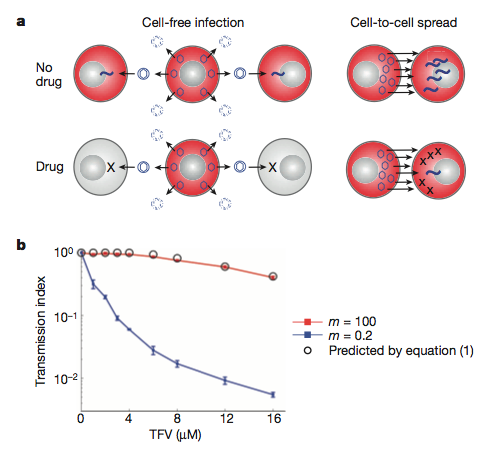
Their model: a) inefficient cell-free infection will be eradicated with antvirals. The more efficient direct cell-cell spread will persist as it involves many more virus particles. b) The mathematical model (backed up by experimental data) showing the loss of infection (transmission index) with increasing concentration of antiviral (TFV) when we have few (m =0.2) or many (m = 100) virus particles.
Expanding the model
Initially, they generated - and experimentally verified - a mathematical model of how HIV transmission may differ during antiretroviral treatment under cell-free (inefficient - low virus transmission) or direct cell-cell spread (efficient - high virus transmission) mechanisms. Their results indicate that when there are many viruses around, the infection is more resistant to the drugs. This, they suggest, is due to the increased probability that at least one virus particle will not interact with the antiviral.
This observation extended to direct cell-cell spread when they introduced previously infected cells with non-infected ones and measured transmission of virus. Both the experimental and theoretical modelling suggest that this spread could be responsible for the ongoing replication seen in patients being treated with antivirals.
What does this mean for HIV therapy? Well, this work brings experimental evidence on the already known process of ongoing HIV replication in the face of antivirals. Through experimentally identifying this mechanism, the researchers here have uncovered a weak-point in HIV biology, one that is not currently being targeted. This method of cell-cell spread may also facilitate escape from neutralizing antibody responses through physically preventing their interaction with HIV proteins. How then might our own bodies combat this virus if its ability to more from cell-cell was inhibited? This work further adds to the growing body of work highlighting direct cell-cell spread as a principle mode of transmission for these retroviruses.
Sigal A, Kim JT, Balazs AB, Dekel E, Mayo A, Milo R, & Baltimore D (2011). Cell-to-cell spread of HIV permits ongoing replication despite antiretroviral therapy. Nature PMID: 21849975
According to WHO data, 2009 saw 33.9 million people worldwide infected with the human immunodeficiency virus - HIV and of these, 1.8 million died from AIDS while 2.9 million were newly infected with the virus. This leaves a year-on-year increase of just over 1 million HIV positive people of which, many of these will go on to pass the virus. Therefore any strategy to eliminate HIV from the human population will have to aim at both treating those already infected as well as preventing new viral transmission and if this is achieved, HIV infection worldwide would dramatically decrease with every year, severely reducing the global health burden caused by this viral pandemic. The only problem is, how can we successfully prevent transmission?
Despite a significant decrease the number of people infected by HIV continues to grow year on year
HIV entry and pathogenesis - where and how can we target it?
One of the major routes of HIV transmission is through sexual activity: an HIV positive person will be carrying many copies of the HIV genome within their cells and in turn these will be able to generate new infectious virus particles. These virions - either present in blood or semen - may be mechanically transferred from infected to uninfected people during sexual activity and many immune cells (Langerhans cells, macrophages and intraepithelial CD4+ T cells to be precise - see below) lining and within the mucosal epithelial surfaces of the reproductive tract are the initial target cells for HIV entry into the human body. It is here that the cells are exposed to the virus through sexual contact, facilitating virus uptake and initial infection then allowing the virus to spread within the body and potentially set up the chronic infection that may develop into AIDS.
HIV entry via immune cells within the vaginal mucosa and spread to systemic lymphoid organs. Miller, 2007.
Many potential strategies are currently in development which aim at preventing person-person transmission during sexual activity through the inhibition of HIV particle transfer. This is the reason why condoms - male and female - are so effective. As the authors of the paper outlined below highlight the requirements of such a strategy:
In many cultural settings, women need a product that can be used covertly without obtaining the permission of their sexual partner. In addition, the cost of HIV prevention must be affordable to the developing world. Thus, there is still a need for products that block HIV transmission, are safe and easy to use, and are coitally independent, discreet, and cost effective.
Bacterial symbiosis to the help
All being so, our bodies are not completely defenseless when it comes to preventing sexually transmitted infections, including HIV; we have multiple tricks up our sleeves and one of which is through a form of bacterial symbiosis. The human reproductive tract is covered in a bacterial biofilm composed of a few species of bacteria, predominantly Lactobacilli. These microbes regulate vaginal biology and aid in the protection against variable infections through the formation of a physical barrier and through alterations in pH. But of course it isn't enough and as I mentioned earlier, people are all too often getting infected with HIV. Yet what if we could enhance these bacterial defenses through genetically engineering those bacteria that natural colonise the reproductive tract? This is exactly what a recently published paper in Mucosal Immunology reports - the development of a novel genetically engineered live bacterial strain expressing an anti-HIV protein that can be easily applied to the vagina and prevents HIV transmission
Normal tissue properties A) and Presence of anti-HIV protein CV-N
The group had previously generated an engineered strain of Lactobacillusjensenii (termed:1153-1666) that expressed and secreted a modified Cyanovirin-N (CV-N) protein. This protein has been shown to have a broad inhibitory activity against a range of HIV-1 strains and provide protection from infection in a non-human primate model of HIV through inhibiting virus entry into those target immune cells - it is also highly potent and non-toxic. To establish the potential for administering this bacteria in humans, the group inoculated the vaginas of non-human primate macaques on a regular basis and achieved stable colonisation along with expression of the antiviral protein in the fluid lining the tract; the recombinant L. jensenii was detected along the epithelium while no tissue changes were observed and no untoward inflammation was detected, all indicating the safety of this strategy.
in vitro HIV inhibition with non-recombinant (left) and recombinant (right) bacteria
So far, the ability of this recombinant bacteria to inhibit HIV infection has not been addressed so later it was assessed through the use of an in vitro model that accurately portrays the physical and biological architecture of the human vagina as well as the macaque model that uses chimeric human/simian immunodeficiency virus infection. Colonization of the in vitro vaginal tract with the non-recombinant bacteria resulted in a 23% reduction in HIV infection compared to the control while inoculation with the CV-N expressing strain gave 72% inhibition (see above). Groups of macaques were then treated with an antibiotic to remove any endogenous Lactobacillus colonizing their vaginal tract and were subsequently inoculated repeatedly with the recombinant strain. These monkeys were then repeatedly challenged with the virus in a manner similiar to what would happen under natural human conditions and the ability of the virus to infect each animal was assessed over time. This strategy reduced the infection rate by nearly 63% (see below).
Macaque challenge outline and results of bacterial colonization protection
This group has generated a bacterial strain - closely related to that already present within the human reproductive tract - that has been genetically augmented through the introduction of the gene for a potent HIV-inhibitory protein Cyanovirin-N. This recombinant bacteria can be inoculated into the vagina and affords protection against initial HIV infection in an in vitro and in vivo macaque model through the expression and secretion of CV-N without generating a toxic response; this engineered microbe is thus better at preventing infection that the non-CV-N expressing bacteria. It remains stable over time But, how then would this function in the real world?
In order to halt the HIV pandemic, we require an at least partially effective strategy to prevent person-person transmission of the virus and this method must safe and inexpensive if it is to be administered to the many people who require it across the developing world. This method outlined above - pending the results of further clinical work - highlights the importance of novel strategies to combat HIV spread. The group outlines the development of an easy-to-apply, safe, efficacious and cheap method to combat HIV transmission in two model systems. Despite these pleasing results, further work will need to be carried out in a clinical field trial to determine whether this mode of protection will work in the real world.
Lagenaur, L., Sanders-Beer, B., Brichacek, B., Pal, R., Liu, X., Liu, Y., Yu, R., Venzon, D., Lee, P., & Hamer, D. (2011). Prevention of vaginal SHIV transmission in macaques by a live recombinant Lactobacillus Mucosal Immunology DOI: 10.1038/mi.2011.30
Through studying viral pathogenesis we seek to understand mechanistically how viral infection and replication causes disease in a particular host. This of course will be subject to a number of complex variables involving both the host and the virus such as: dose; genotype; virus receptor distribution of host tissues;the ability of the virus to replicate in those infected cells and the hosts response to that infection.
Although it is an extremely complex system, knowledge of it may allow us to develop certain preventative strategies alongside new treatments and therapies. That is why being able to study viral pathogenesis is important and why some may welcome a recently published paper reporting initial data (possible cell tropism and host immune response) from a mouse model of infection with xenotropic murine leukemia-related virus (XMRV), a possible novel human pathogen (and a close relation of natural mouse retroviruses).
[caption id="" align="aligncenter" width="258" caption="Mus musculus - relative of Mus pahari. Just incase you forgot what a mouse looked like."][/caption]
Originally identified in a number of human prostate tumour samples, XMRV has since had a conflicting scientific history (covered much better elsewhere), with some studies showing a link between infection and chronic fatigue syndrome (CFS) and prostate cancer. Others since have failed to detect such a link. Despite this, knowledge of how this virus could potentially interact with the human body would of course be useful to acquire.
This understanding has been blocked somewhat by the lack of a small-animal model – a cheap, easier and more ethical alternative to non-human primate studies and of course easier to study than humans. XMRV just does not infect normal lab mice (Mus musculus) and thus pathogenesis in this host does not occur and if pathogenesis doesn’t occur, we can’t study pathogenesis. This species of mouse doesn’t express the receptor that XMRV utilizes to gain entry into cells. Sakuma et al have therefore used Mus pahari, a wild asian relative which does express the receptor for XMRV and thus may tell us something about XMRV pathogenesis.
The group showed that XMRV was able to successfully infect M. pahari cells in vitro and also was able to infect M. pahari following injection of the virus into whole-animals. Following infection, they were able to screen mouse cells and tissues (such as blood) for the telltale signs of XMRV infection over 12 weeks; XMRV being a retrovirus, integrates a DNA copy of its RNA genome into host cells and PCR detection of this integration may allow us to infer XMRV infection. They also investigated the possible role of infectious virus being present in the animal and the of XMRV replication on host cell functioning.
Detection of XMRV DNA in particular tissues may allow us to infer how infection proceeds within the host and how it causes disease. Viral sequences were detected in blood cells, heart, spleen, brain, testis and prostate tissues, although detection was highly variable between mice and a clear picture of infection didn’t really emerge over any of the time periods. The group focused on the effect of XMRV on lymphocytic cell functioning (possibly a good place to start giving its apparent involvement in CFS, an immunological disorder): CD4+ T helper cells and CD19+ B cells being targeted within the spleen. Over all, in some of the mice, increased total white-blood cell numbers was observed early in infection indicating a possible deregulation of lymphocyte development, although this was by no means common and only slight.
The role of both mouse adaptive and innate immunity was also assessed and interestingly, the mice generated a robust antibody response to XMRV antigens yet no long-term studies were involved to see how the virus adapted. Viral genome sequences found within blood and spleen tissues were sequenced and those from the spleen only, displayed predominant G-to-A hypermutation possibly indicative of intra-tissue restriction of viral replication. Host-mediated mutation of viral genomes will therefore more likely result in highly defective viral sequences and possibly prevent future viral infections; one example is that of APOBEC3-like enzymatic reactions.
Of course, these data are rather preliminary with optimisation of infection expected to come later, but at the minute, how does this relate to what happens in humans and will this model actually be on any use?
The development of a permissive small-mammal model of XMRV infection described here will certainly facilitate scientific investigation as it has done for many other viruses although it must be remembered that what happens in a mouse may not happen exactly that way in humans.
This study, however does appear to shed light on cell tropism of XMRV and possibly its transmission - blood-borne (although this was looked at really).
The possible deregulation of host lymphocyte development may play a role in the pathologies associated with it in humans. The highly variable pathological outcomes may be due to the relatively less homogenous gene pool of M. pahari rather than anything to do with the virus - although this may be occurring.
The fact that these mice developed strong antibody responses to this virus may allow the development of vaccine strategies.
But, of course, the importance and relevance of this work will all come down to whether it is or is not a true human pathogen yet we will certainly benefit from this work if it turns out that it is. Expect a lot more from this group if it is.
Sakuma T, Tonne JM, Squillace KA, Ohmine S, Thatava T, Peng KW, Barry MA, & Ikeda Y (2011). Early Events in Retrovirus XMRV Infection of the Wild-Derived Mouse Mus pahari. Journal of virology, 85 (3), 1205-13 PMID: 21084477
Sakuma T, Tonne JM, Squillace KA, Ohmine S, Thatava T, Peng KW, Barry MA, & Ikeda Y (2011). Early Events in Retrovirus XMRV Infection of the Wild-Derived Mouse Mus pahari. Journal of virology, 85 (3), 1205-13 PMID: 21084477




 [/caption]
