Field of Science

Showing posts with label non-human primates. Show all posts

Can we prevent gorilla extinction with vaccination?

Western Lowland gorilla
Gorillas, Gorilla spp. are found only throughout central African rainforest where there are in total over 200,000 individuals living in the wild. Two gorilla species are recognised, split between east and west Africa with at least two sub-species recognized in both. Their numbers are rapidly decreasing with problems such as habitat loss, poaching and human war contributing  greatly to a rapid reduction in their numbers. Sadly, one other factor on top of these in which these large primates must worry about is that of the transfer of infectious agents arising from humans and other animals. 

Viruses are constantly being transferred between populations of animals but may not establish infection all that easily in a non-host species. Although occasionally infection will result in virus replication and significant disease and this is known as a zoonotic infection -  when non-human viruses are transmitted to humans (see HIV, Influenza A and SARS-coronaviruses) and 'reverse zoonosis' when human viruses infect other animals. This pathogen transfer may be especially important when occurring in a critically endangered species such as gorillas; one recent example is that of human metapneumovirus.

EBOV effects not just humans http://turbo.indyposted.com/

We have been able to detect the effect of a number of viruses on gorilla populations including enteroviruses, adenoviruses and parvoviruses for example, although a small number are known to cause disease. In the last decade thousands of gorillas, chimpanzees and other mammals were killed through infection with the Zaire strain of ebola virus (EBOV) in the rainforests throughout the Congo basin area of central Africa. EBOV is a highly infectious and deadly RNA filovirus which causes a nearly always fatal hemorrhagic fever. The reservoir species for EBOV has been linked to central African fruit bat populations and ebola has caused hundreds of human deaths since its first recorded emergence in the 1970s.  Significantly more non-human primates have been victim to ebola than humans. We can implement a number of control measures for example limiting human-ape contact especially when ill to prevent this virus transfer but this may be more difficult when humans are not involved as in the case of EBOV. There are also a number of therapeutic options available although in the case of ape infection, would be logistically impossible. One strategy we therefore must consider is that of potentially protecting these Gorilla populations through vaccination against a number of potential viral pathogens.

The group VaccinApe is attempting to do just that. A volunteer consortium lead by the charity group the World Wildlife Fund, a vaccine developer Integrated Biotherapuetics and two academic institutions, the Max Plank institute for Evolutionary Anthropology and Kansas state University, VaccinApe is trying to develop an easy and effective method of vaccinating Gorilla populations in the wild. Currently in a 'proof of concept' phase, the group will lead the development of non-human primate vaccinology in order to generate a safe and reliable Ebola virus vaccine to be used through darting of individual gorillas. A large scale vaccination program may therefore afford protection of critically endangered gorilla populations against future EBOV emergences.

In light of the clinical severity of EBOV infection in humans a number of potential vaccine candidates have been developed which rely upon the generation of a protective immune response specifically to EBOV. One main candidate the group are interested in is the EBOV virus-like particles (VLPs). These are effectively non-infectious viruses lacking a viral genome but retaining virus antigenic proteins. Therefore EBOV-specific immunity will be generated against whatever EBOV proteins are found within the VLP. In early trials in macaques, this VLP strategy protected individuals against EBOV challenge although whether this could safely be transferred to gorillas isn't known.


EBOV VLP. Looks and acts antigenically like 'live' ebola. http://www.integratedbiotherapeutics.com/

Endangered wild gorilla and chimpanzee populations are at a great risk from a number of emerging viruses with the most important being EBOV. The difficulties in preventing direct transmission/therapeutic intervention have led people to consider the development of anti-EBOV vaccines. A number of candidates have already been tested and proved safe and effective in non-human primate models possibly allowing these vaccines to be transferred to gorilla population testing. It is hardly surprising that the work required to carry out such large-scale and difficult vaccination campaigns in wild gorillas in the African rain forest will be extremely difficult. The main problems include safety/efficacy testing in gorillas, physical vaccination methods and tracking anti-EBOV immunity non-invasively. Despite these difficulties, only time will tell whether the work of VaccinApe and their partners is to be supported as a worthwhile investment to save these animals.

ResearchBlogging.org

Le Gouar PJ, Vallet D, David L, Bermejo M, Gatti S, Levréro F, Petit EJ, & Ménard N (2009). How Ebola impacts genetics of Western lowland gorilla populations. PloS one, 4 (12) PMID: 20020045


Richardson JS, Dekker JD, Croyle MA, & Kobinger GP (2010). Recent advances in Ebolavirus vaccine development. Human vaccines, 6 (6), 439-49 PMID: 20671437

Warfield KL, Swenson DL, Olinger GG, Kalina WV, Aman MJ, & Bavari S (2007). Ebola virus-like particle-based vaccine protects nonhuman primates against lethal Ebola virus challenge. The Journal of infectious diseases, 196 Suppl 2 PMID: 17940980

So, how do you know when a vaccine is safe?

How can you tell how safe a vaccine is?
ResearchBlogging.org
Mumps, a highly infectious viral disease, has been largely eradicated in the developed world following the introduction of a highly effective live-attenuated vaccine. Highlighted by well-publicized outbreaks in the U.S and U.K, the number of cases, however, has risen causing worldwide alarm. The reasons for this re-emergence have yet to be fully elucidated but most likely are due to a number of factors, including waning immunity and poor vaccine coverage.

Despite what is normally reported, mumps infection can cause serious complications. Prior to the introduction of the vaccine - and of course in countries that fail to administer it - mumps infection was/is the most common cause of viral meningitis and encephalitis; it has been estimated that 50% of those infected by mumps have some form of central nervous system involvement, although 1 - 10% will actually experience a symptomatic infection. It is safe to say that the mumps virus is one of the most neurotropic human viruses currently circulating and that its neurotropism can hardly be considered a complication.

All this really underlines the importance of maintaining mumps vaccination in protecting individuals and populations from serious disease. The key then is to develop not just more effective vaccines but also safer vaccines as people aren't likely to give their children a vaccine which may cause serious side-effects especially considering the propensity for mumps virus to cause CNS disease. A recent review of mumps vaccine safety states that,

Such a problem places public confidence in all mumps vaccines at risk, as indicated by the experience in Japan where national mumps vaccination programs were discontinued in 1993 following established links to aseptic meningitis; consequently, more than a million new mumps cases occur annually in that country


Lewis rat - is this the future of mumps vaccine safety?
How then are we to assess the safety and more specifically 'neurovirulence' of mumps candidate vaccine stocks? Recently, Rubin and Afzal from the United States Food and Drug Administration and the UK National Institute for Biological Standards and Control respectively, outlines the current state of the art in mumps virus safety testing and outlines how its future might look. What we would like in a test system is for it be accurate and fully predictive (limit false positives and negatives); it would need to economical (vaccines need a lot of testing) and it needs to be relatively easy to carry out and replicate. For us to do this, these methods require vigorous testing!

Currently, much like other virus vaccines, mumps vaccine safety is assessed in a monkey model and has resulted in the detection of significantly attenuated vaccines for use in humans. There is however cause for concern with this system as in some instances it fails to distinguish between important differences in levels of attenuation. There is therefore a need to replace this system if not on the grounds of ethical and economic concern but on the grounds of safety. In has stepped a small animal model - the lewis rat- which has been shown to better predict neurovirulence; is cheaper and is less ethically taxing; it is hence subject to a WHO validation study.

False colour electron micrograph of the mumps virus

But why do we have to use animal models at all for safety testing? Can we not just be content with in vitro studies with cell lines? In some cases, we can predict how a virus will act within an animal on the basis of studying how it infects and replicates in cell line but there is, however, no in vitro alternatives for mumps - at least not yet - and even if there were we can't say whether it could ever fully replace animal studies.

In some systems, animal infections just cannot be replaced if we are to maintain a high level of vaccine safety which of course is important when vaccines are administered to billions of people worldwide we are then forced to stick with animal testing. We can rest assured that with recent developments in small-animal models, future testing may come more accurate, cheaper and a little more ethically pleasing.

BRUYN HB, SEXTON HM, & BRAINERD HD (1957). Mumps meningoencephalitis; a clinical review of 119 cases with one death. California medicine, 86 (3), 153-60 PMID: 13404512

Dayan GH, & Rubin S (2008). Mumps outbreaks in vaccinated populations: are available mumps vaccines effective enough to prevent outbreaks? Clinical infectious diseases : an official publication of the Infectious Diseases Society of America, 47 (11), 1458-67 PMID: 18959494

Rubin, S., & Afzal, M. (2011). Neurovirulence safety testing of mumps vaccines—Historical perspective and current status Vaccine DOI: 10.1016/j.vaccine.2011.02.005

Can fluorescent-‘labelled’ viruses illuminate their mechanisms of pathogenesis?

Have you ever wanted to visualise viral infection? Ever wanted to observe how they enter and spread throughout their host organism? Ever wanted to know how exactly they caused disease - at the cellular and whole-organism level? Well, this may be entirely possible using fluorescent-labeled recombinant viruses infecting a relevant model system.

[caption id="" align="aligncenter" width="504" caption="GFP-virus infected cells"][/caption]

So how does it work?

Lemon et al recently report the continued investigation of measles virus pathogenesis in a non-human primate (Macaque) model utilising a green-fluorescent protein (GFP) expressing virus. Upon infection of host cells, viral transcription leads to the very high expression of GFP, flooding the cytoplasm with this fluorescent ‘tag’. Subsequent microscopy, imaging and immunohistochemistry allows for the identification and location of the infected cells, tissues and organs - see image above. Tracking of cellular infection allows us to decipher the development of MeV entry, spread and replication at both the cellular and whole-organism level throughout the entire infection. Studies such as these give an unprecedented view of viral infection in a means directed related to that of human infection. This model even allows for macroscopic real-time detection of fluorescence and hence viral infection.

Why is this important for measles?

Despite a highly effective vaccine and significant global control initiatives, measles infection still accounts for significant morbidity and mortality worldwide, mostly in the developing world (164,00 deaths in 2008). This is mostly attributable to the profound immunosuppression induced allowing for further infection with opportunistic pathogens. Currently, much is known about measles pathogenesis yet the molecular mechanisms of such are poorly understood and it is therefore of great interest to better understand these processes by which MeV infects and causes disease in humans. Knowledge of such may facilitate the development of more effective and safer vaccines for measles and indeed other viral pathogens.

Viruses being obligate intra-cellular parasites, must enter and exit cells in order to survive. Most of viral pathogenesis can therefore be attributed to the effects of viral replication of host cells and tissues; a major determinant of which is the expression of receptors on host cells surfaces allowing viral entry, infection and replication. Currently only a single receptor – CD150 - (otherwise known as signalling lymphocyte activation molecule SLAM) has been discovered that wild-type pathogenic MeV uses to enter host cells; the distribution of which only explains part of measles pathogenesis as epithelial and neuronal cells (important target cells) do not express the protein. As indicated by this receptor being expressed on lymphocytes and other immune cells, MeV is a highly lymphotropic virus! But if epithelial cells fail to express the receptor on their surface, how come its possible for MeV to enter via these cells?

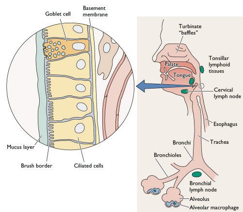
The classical view of measles pathogenesis was that free-virus entered the host through the respiratory route, infecting and primarily replicating within the epithelial cell lining of the respiratory tract. Newly produced virus spreads to nearby lymph nodes where infected monocytes – a type of immune cell - facilitates viral dissemination throughout the host, resulting in the well-known symptoms of measles. The problem with this being that epithelial cells and unstimulated monocytes fail to express the MeV receptor CD150 and infection should therefore not occur. Recently, it has been shown (again using a GFP expressing virus in a macaque model) that MeV predominately infects dendritic cells during the peak of infection, ruling out a major role for monocytes. There is also however no direct evidence of MeV primary replication within the epithelium of the respiratory tract at the early stages of infection. So what exactly happens during the start of infection and does it develop? GFP-expressing viruses may shed light on this question.

[caption id="" align="aligncenter" width="415" caption="Diagramatic representation of the cellular composition of the human respiratory tract - notice the epithelial cell lining and the alveolar macrophages. Dendritic cells are however not shown on this diagram."]Diagramtic representation of the cellular composition of the human respiratory tract - notice the epithelial cell lining and the alveolar macrophages. Dendritic cells are however not shown on this diagram.[/caption]

So how can we study the early stages of infection?

The incubation period of  measles is about 2 weeks in humans making it particularly difficult to study the early events of viral infection – the kind of events like host entry, initial site of replication and subsequent intra-host dissemination - this is where we can use a non-human primate model.

Lemon et al  generated a highly virulent recombinant MeV based on viral isolates from an outbreak in Sudan; they engineered the viral genome so that it expressed GFP upon entry into cells – an addition that causes little or no replication defects to the virus. Groups of macaques were subsequently infected via the respiratory route allowing highly sensitive visualisation of GFP expressing cells following necropsy. The early time-points of around 5 days post infection were focussed on in this investigation allowing the determination of the early cell targets - epithelium? Immune cells?

So what did they find?

Their results suggest that at the early stages of MeV infection, GFP and hence viral replication is only found in immune cells within the respiratory tract and not the epithelial lining. Dendritic cells and alveolar macrophages are believed to capture viral particles in the lungs allowing spread via infected cells. This is known as a Trojan horse entry mechanism like that used by HIV to pass through mucosal tissues and infect humans - see below. This infection allows for spread and localised replication within nearby lymphoid tissues and then on to draining lymph nodes where massive lymphocyte cell infection may occur facilitating dissemination throughout the host, mainly within lymphoid tissues. Virus can be carried through host blood vessels to other lymphoid target tissues like the tonsils and adenoids and the gut-associated lymphoid tissue ' Peyer's patches'.

[caption id="" align="aligncenter" width="465" caption="HIV entry mechanisms utilising dendritic cells to pass through epithelial cell barriers - the 'Trojan horse' mechanism. This may be directly analogous to MeV entry and primary spread except in the respiratory tract."][/caption]

What does this mean?

This study clearly demonstrates the importance of non-epithelial cells such as dendritic cells in MeV entry, early replication and subsequent systemic spread. It does not however, rule out a major role for epithelial cells in later stages and in transmission - MeV still infects non-CD150 expressing cells and currently the mechanisms of which are unknown. Focusing on the later stages of infection may allow us to appreciate the other cell targets in pathogenesis and viral transmission. As mentioned previously, the use of fluorescent-labeled viruses offers an unprecedented view of viral entry, spread and pathogenic mechanisms. We should look forward to the time when studies like these are applied to other viral and indeed non-viral pathogens.

ResearchBlogging.orgLemon, K., de Vries, R., Mesman, A., McQuaid, S., van Amerongen, G., Yüksel, S., Ludlow, M., Rennick, L., Kuiken, T., Rima, B., Geijtenbeek, T., Osterhaus, A., Duprex, W., & de Swart, R. (2011). Early Target Cells of Measles Virus after Aerosol Infection of Non-Human Primates PLoS Pathogens, 7 (1) DOI: 10.1371/journal.ppat.1001263

Coombes, J., & Robey, E. (2010). Dynamic imaging of host–pathogen interactions in vivo Nature Reviews Immunology, 10 (5), 353-364 DOI: 10.1038/nri2746

Uncovering the biomedical and evolutionary importance of primate innate immunity


Primates such as chimpanzees differ in the diseases that affect them - especially when we compare them to those that affect us. Progression to AIDs, cancer incidence, Alzheimers disease and malaria either do not affect or cause less severe diseases in non-human primates than they do in humans. So why are our primate cousins so differently affected by these particular diseases than we are? What causes these differences? Are they environmentally mediated? Behavioural? Molecular?

What does the innate immune system have to say about it?

The pathogenesis of disease is an important area of research when trying to understand the origins and progression of diseases. It depends on many variables - host specific, environmental or pathogen specific. A recent publication has sought to understand these differences from the point of view of host factors mainly the innate immune system. By comparing genome-wide gene expression patterns in primary immune cells (monocytes) cultured in vitro from groups of humans, chimpanzees and rhesus macaques and applying bioinformatic analysis to the results, the investigators were able to detect those genes whose expression is altered upon stimulation with lipopolysacharide (LPS), an important activator of the innate immune system. From this data set they could ask, functionally, what makes the human immune system different?


3,170 genes were seen to be differentially expressed with 793 changing in all three species indicating a conserved function. Other genes showed species-specific changes allowing researchers to ask what makes each species unique? More importantly what makes non-human primates different when it comes to diseases? There were 335 genes in the human monocytes that were expressed differently and these were divided between particular pathways.

What does this data have to say about specific diseases?

Those genes listed as being involved in viral infection were those most likely to be species-specific indicating the rapid adaptation of host immunity to fast-evolving viral pathogens. The data sheds light on the possible lo incidence of cancer in non-human primates by detecting the difference in apoptosis/cancer related genes - although the signifigance of monocyte gene expression when considering the whole-organism in diseases such as cancer is difficult to say. Most interestingly seen are those genes involved in HIV infection and AIDs  whose expression may explain why chimpanzees do not progress to AIDs or do so slowly.

Despite the problems in culturing non-human primate immune cells in vitro and the difficulties in controlling for environmental effects (different diets of primates), this study goes some way to understanding at a functional level what makes humans human from an immunological point of view. Although focussing on a single cell type, the monocyte, other cells may prove useful in investigating innate differences - as other cells also function in innate immunity - mucosal epithelium for one. This work paves the way for more detailed molecular analysis but also of more genome-wide work looking at other cells, other activators and pathogens (not LPS but HIV?). Understanding what makes humans different in a pathogenic light should focus not just on immunology at the gene expression level but also differences in epigenomics, behaviour, anatomy and cell biology.


Varki, Aijit. 2000. A Chimpanzee Genome Project is a Biomedical Imperitive. Genome Res. 2000. 10: 1065-1070. doi: 10.1101/gr.10.8.1065

Barreiro, Luis B., John C. Marioni, Ran Blekhman, Matthew Stephens, and Yoav Gilad. 2010. Functional Comparison of Innate Immune Signaling Pathways in Primates. Ed. Greg Gibson PLoS Genetics 6, no. 12 (December): e1001249. doi:10.1371/journal.pgen.1001249. http://dx.plos.org/10.1371/journal.pgen.1001249.