There are currently two AIDS pandemics raging across the world: the well-known one in humans and the less recognised one in domestic cats. Feline AIDS, caused by the feline immunodeficiency virus (FIV) - a close relative to HIV - is pretty similar to that afflicting millions of humans and could potentially be used as a model system for studying HIV infection and disease. One major scientific barrier to HIV research in primates is the difficulty in generating transgenic animals efficiently. This, however, could be sidestepped through employing FIV/domestic cat as the experimental system.
The 9,000 year-long relationship between the human species and domestic cats has brought a lot to each respective species: we get rid of nasty pests and enjoy the whole 'cat experience' while they feast on the human-associated rodents and gain some shelter and people love. But what some people might not realise is that the domestic cat has - and will probably continue - to help us in biomedical research. And, for them, the increased research into cat biology may also spill over into non-domestic species, who are on the brink of extinction and might be aided by this work.
The sheer amount of genomic data now available from a wide range of species has allowed the increased scrutiny over what genes and DNA sequences are present in their chromosomes. What we have begun to notice is that many of these sequences have a viral origin.
And, in the recent half-decade, the numbers of these endogenous viruses discovered have rapidly increased, but how did they get there? What are they doing? And, are they bad for us? Only a true experimental model system can answer these question but this is something which is lacking.
Lets talk about ERVs
Now, viruses have left their mark on our genomes in more ways than one; infection and associated disease/mortality has heavily influenced the genetic structure of populations via natural selection and genetic drift for millions of years. Yet, another important mechanism is that employed by the endogenous retroviruses (ERVs) that have inserted a DNA copy of themselves into our chromosomes - the norm for retroviruses - and have forever become part of us.
Over the course of evolution, these once infectious viruses have become redundant, building up a collection of genetic mutations resulting in loss of replicative ability. Although many still play a role in the cellular biology of the host and have been a great source of genetic novelty over the billions of years of evolution.
However, what we have noticed is that viruses other than retroviral species have inserted themselves into genomes of humans, other animals and even plants and fungi. Many of these viruses have a DNA phase in their replication cycle, which is put into the genome of their host to aid their survival and so it may not be all that surprising that they have stayed with us through evolution (these viruses include many single-stranded DNA viruses and again).
One intriguing observation is that many of these non-retroviral endogenous viruses are in fact - or were - RNA viruses with no known DNA phase during replication. They are therefore called Non-retroviral RNA virus sequences (NRVSs). See plant NRVSs and mammalian NRVS (ebola virus-like , borna virus-like and many more - (lots, they're everywhere). There is strong evidence that these integrations occurred thousands, if not millions of years ago and could have played a role in the evolution of many species.
How can we study these viruses?
But just exactly how do these viruses do it? After-all, they are RNA viruses without a reverse-transcriptase enzyme and hence no natural ability to produce a DNA genome that can be inserted into our chromosomes. And, can we follow this endogenisation experimentally? One mechanism is thought to occur when an endogenous retrovirus-like element joins itself to a non-endogenous RNA virus and then this chimera is put into our genome. But this is really only half the story - can we ever study the entire process, from initial infection to endogenisation?
For an RNA virus to become fully integrated into our germline it has to first infect our germ-line cells (sperm/oocytes); its RNA genome must be copied into DNA and this DNA molecule must be inserted into the chromosome. It also must allow for the development of healthy and reproductively active offspring and can then let evolution take its course. An experimental model system of this process would allow for a better understanding of this process in molecular detail and how this relates to the evolutionary process as a whole.
Here's how you would do it:
The animal model
Bank vole - a good model for endogenous viruses?
A small-animal model that could be infected by a type of virus that had been shown to integrate into the genome (borna disease virus, for example) would make this easier to study. Plus, many rodents have been shown to harbour many NRVSs already.
The virus infection
You would infect the animals with the virus in as natural conditions as possible and look to see whether the virus entered and replicated in the cells of the germ-line.A GFP-expressing virus would work best for this.
Detection of RNA - DNA
What you would have to do is be able to track the process of turning the RNA genome into DNA. A PCR-based screening would work well for this and could be applied to a range of tissues in the host, including occytes/spermatozoa.
Integration
To prove that the DNA copy was inserted into the host chromosome you would need to sequence the sites where the DNA had integrated in and determine where in the genome it lay.
Stability
This experimentally infected rodents could be bred continously and the presence of endogenised virus looked for in their offspring. The expression of said virus genes (if there is any) could be followed in rodent tissues.
Borna disease PCR without reverse-transcriptase. A) no nuclease treated, B) RNA nuclease treated, C) DNA nuclease treated and D) PCR with reverse transcriptase step
Well one paper has maybe taken the first step in the development of such a model system (although they may not know it). It has shown evidence that if you infect baby bank voles with borna virus, directly into their brain you can detect borna virus-specific DNA sequences using PCR following DNA extraction (see above PCR gel for results). And, these sequences resulted from the virus, not some already-endogenised borna virus sequence. Although they did not check for germ-line infection or integration, this is the first step. The applicability of Borna virus reverse genetics and these animal models could make this kind of study feasible but certainly not easy. We may in future catch a glimpse of this process in real-time.
Kinnunen, P., Inkeroinen, H., Ilander, M., Kallio, E., Heikkilä, H., Koskela, E., Mappes, T., Palva, A., Vaheri, A., Kipar, A., & Vapalahti, O. (2011). Intracerebral Borna Disease Virus Infection of Bank Voles Leading to Peripheral Spread and Reverse Transcription of Viral RNA PLoS ONE, 6 (8) DOI: 10.1371/journal.pone.0023622
Mumps, a highly infectious viral disease, has been largely eradicated in the developed world following the introduction of a highly effective live-attenuated vaccine. Highlighted by well-publicized outbreaks in the U.S and U.K, the number of cases, however, has risen causing worldwide alarm. The reasons for this re-emergence have yet to be fully elucidated but most likely are due to a number of factors, including waning immunity and poor vaccine coverage.
Despite what is normally reported, mumps infection can cause serious complications. Prior to the introduction of the vaccine - and of course in countries that fail to administer it - mumps infection was/is the most common cause of viral meningitis and encephalitis; it has been estimated that 50% of those infected by mumps have some form of central nervous system involvement, although 1 - 10% will actually experience a symptomatic infection. It is safe to say that the mumps virus is one of the most neurotropic human viruses currently circulating and that its neurotropism can hardly be considered a complication.
All this really underlines the importance of maintaining mumps vaccination in protecting individuals and populations from serious disease. The key then is to develop not just more effective vaccines but also safer vaccines as people aren't likely to give their children a vaccine which may cause serious side-effects especially considering the propensity for mumps virus to cause CNS disease. A recent review of mumps vaccine safety states that,
Such a problem places public confidence in all mumps vaccines at risk, as indicated by the experience in Japan where national mumps vaccination programs were discontinued in 1993 following established links to aseptic meningitis; consequently, more than a million new mumps cases occur annually in that country
Lewis rat - is this the future of mumps vaccine safety?
How then are we to assess the safety and more specifically 'neurovirulence' of mumps candidate vaccine stocks? Recently, Rubin and Afzal from the United States Food and Drug Administration and the UK National Institute for Biological Standards and Control respectively, outlines the current state of the art in mumps virus safety testing and outlines how its future might look. What we would like in a test system is for it be accurate and fully predictive (limit false positives and negatives); it would need to economical (vaccines need a lot of testing) and it needs to be relatively easy to carry out and replicate. For us to do this, these methods require vigorous testing!
Currently, much like other virus vaccines, mumps vaccine safety is assessed in a monkey model and has resulted in the detection of significantly attenuated vaccines for use in humans. There is however cause for concern with this system as in some instances it fails to distinguish between important differences in levels of attenuation. There is therefore a need to replace this system if not on the grounds of ethical and economic concern but on the grounds of safety. In has stepped a small animal model - the lewis rat- which has been shown to better predict neurovirulence; is cheaper and is less ethically taxing; it is hence subject to a WHO validation study.
False colour electron micrograph of the mumps virus
But why do we have to use animal models at all for safety testing? Can we not just be content with in vitro studies with cell lines? In some cases, we can predict how a virus will act within an animal on the basis of studying how it infects and replicates in cell line but there is, however, no in vitro alternatives for mumps - at least not yet - and even if there were we can't say whether it could ever fully replace animal studies.
In some systems, animal infections just cannot be replaced if we are to maintain a high level of vaccine safety which of course is important when vaccines are administered to billions of people worldwide we are then forced to stick with animal testing. We can rest assured that with recent developments in small-animal models, future testing may come more accurate, cheaper and a little more ethically pleasing.
BRUYN HB, SEXTON HM, & BRAINERD HD (1957). Mumps meningoencephalitis; a clinical review of 119 cases with one death. California medicine, 86 (3), 153-60 PMID: 13404512
Dayan GH, & Rubin S (2008). Mumps outbreaks in vaccinated populations: are available mumps vaccines effective enough to prevent outbreaks? Clinical infectious diseases : an official publication of the Infectious Diseases Society of America, 47 (11), 1458-67 PMID: 18959494
Rubin, S., & Afzal, M. (2011). Neurovirulence safety testing of mumps vaccines—Historical perspective and current status Vaccine DOI: 10.1016/j.vaccine.2011.02.005
One worthwhile way to study viruses – and other micro-organisms – is to see where exactly they are found within a host. How do they enter the body? What organs do they infect and how? How do they spread from tissue to tissue and organ to organ? How do they exit the body? These are just some of the questions which it would be good to actually SEE how and where it happens. Maybe then we could better understand the dynamic relationships governing infection and disease; maybe then we could design better, more effective therapies. Just maybe. Like it is all that easy.
How could we track adenovirus movements?
For one thing viruses are pretty small - so, are there any high resolution methods of looking at infection that may be able to help us? Pathologists can look at tissue samples from living or dead patients (animals included) and microscopically assess these for signs of viruses using for example: antibodies specific for particular proteins or nucleic acid probes (in situ hybridisation or PCR perhaps) found only in infected cells. This stuff is pretty good and is routinely used for both, clinical sciences and biological research but its kind of limited and in the last decade or so, the use of ‘reporter genes’ - like GFP and luciferase has facilitated the easier and faster analysis of viral infection.
Reporter genes are inserted into the viral genome and when expressed upon cell infection, they produce a protein – maybe a fluorescent or luminescent protein – whose function can be assayed and followed. This is what we can look at during infection. There are however certain limitations with this, in that, only those viruses that successfully infect a cell will express the reporter gene; viruses which fail to infect and are taken up by the liver or immune cells will not be detected. We are seeing a biased image of viral infection if we only consider reporter gene expression. This is particularly important when we are using viruses as therapeutic agents themselves as strict pharmacological testing requires intimate details of in vivo distribution and kinetics. So how can we see viruses in vivo without reporter genes?
As a paper in PLoS ONE, from a group at the Mayo Clinic in Rochester, demonstrates, there IS an alternative way to track viruses in vivo – the molecular attachment of individual reporter molecules to the virus itself thus requiring no gene expression and therefore allowing a more unbiased view of infection. An interesting aspect of this work is that it is carried out in real-time; these reporter molecules can be visualised as infection happens, at the millisecond scale. This also allows for the tracking at very early time points, times where no viral gene expression is taking place.
This study came at it from the angle of developing safer and more effective anticancer viruses, viruses which will infect and kill only those malignant cancer cells but this could be applied to any area of investigation. Being able to follow virus distribution in a mouse-model is of course a great scientific and clinical benefit to them. The group dyed an adenovirus vector with a molecule which emits light in the near-infrared range (particularly suited to in vivo imaging) which they then injected into groups of mice via their jugular vein. They were thus able to analyse and quantify the tissue distribution of their labelled vector throughout a whole mouse, in real-time.
This is the first time that this kind of imaging has been carried out and it certainly won’t be the last. This work could be applied to yet more viral vectors; it could be used to study a ‘natural’ infection or it could be used for non-viral imaging of therapeutic nanoparticles. A combination of this early time-point analysis with later, reporter gene expression imaging would be able to give us an unprecedented view into dynamic real-time viral infections in a number of model systems.
Hofherr, S., Adams, K., Chen, C., May, S., Weaver, E., & Barry, M. (2011). Real-Time Dynamic Imaging of Virus Distribution In Vivo PLoS ONE, 6 (2) DOI: 10.1371/journal.pone.0017076
Despite the development of effective vaccines, many human populations are currently at the mercy of numerous endemic viral pathogens. Measles virus is one such pathogen that, in 2008, was responsible for 164,000 deaths; the worst effected areas are South-East Asia and Africa (WHO stats can be found here). You might find this surprising as there is currently a very good measles vaccine in use – in fact you probably received at some point during childhood and are protected from future infection. Measles cases have been significantly reduced in the developed world, so why hasn’t this vaccine allowed for the eradication of measles virus transmission in the developing world?
Needle vaccination against measles
The key to controlling measles – and other viruses – is to generate sustained high levels of good quality immunity within a population so that the virus can no longer successfully infect and has nowhere to go; this is known as herd-immunity. The problem then is, well why can’t we achieve the herd immunity required to prevent virus transmission? In places like Africa, where people are reminded daily of the horror of measles, you don’t have to force them to accept vaccination unlike what was seen in the UK and US recently so they are readily vaccinated. One problem however, appears to be the mode of vaccination, that is injecting the virus vaccine creates hurdles to a successful immunisation campaign:
·Trained healthcare workers are required to safely administer the vaccine when it is injected
·The currently used vaccine formulation tends to go off in temperatures ~ 37 degrees Celsius causing problems for transport and storage especially in areas such as Africa.
·The use of used/contaminated needles may facilitate the problems of blood-borne diseases and drug use
Currently, some 3million children have already been successfully vaccinated from measles using a ‘wet’ aerosol delivery system, however the formulation was unstable above 4 degrees Celsius and delivery was difficult. A recent paper published in PNAS has sought to improve upon this current measles vaccination technology (and also get around the problems of injecting vaccines) through the generation of a highly immunogenic respiratory-delivered ‘dry’ vaccine formulation. It was tested in macaques, can be administered as a single dose and is a highly thermostable, powdered formulation.
'PuffHaler' aerosol delivery system for 'dry' vaccines
So, how good is it?
They report that their aerosol-delivered vaccines were deposited into the upper and lower respiratory tracts and resulted in the generation of good-quality measles virus specific humoral (B cell + antibody) and cellular (CD8+/CD4+ T cells) immune responses without safety concerns; there also exists a long-lived (<1 year) B cell memory function (and some T cell memory) correlating with long-term virus protection. They show the their vaccine strategy allows for the successful protection from subsequent measles virus challenge. Through comparisons with injected vaccine, the group were able to show that indeed all routes of vaccination generated the required level and quality of immunity required to protect from measles but given other concerns with injecting vaccine aerosol delivery may prove better. These results indicate that this proof of concept, novel vaccine may be comparable to the previously used formulation – although human studies would have to be carried out.
Aersoal delivery system
Scientists are being allowed to investigate problems like these only through being funded by the 'Grand Challenge in Global Health Grants' via the Bill and Melinda Gates Foundation; the money supplied allows for the development of better, safer and ultimatey more cost-effective vaccines. This work highlights the importance in the development of these new and more effective vaccine technologies in order to facilitate the eradication of viral pathogens worldwide. How might this method of administration affect other vaccines, only further work will decide but if we are unable to prevent measles transmission with a highly effective vaccine then what hope do we have to prevent other, less well-studied viruses without decent vaccines?
Lin, W., Griffin, D., Rota, P., Papania, M., Cape, S., Bennett, D., Quinn, B., Sievers, R., Shermer, C., Powell, K., Adams, R., Godin, S., & Winston, S. (2011). Successful respiratory immunization with dry powder live-attenuated measles virus vaccine in rhesus macaques Proceedings of the National Academy of Sciences DOI: 10.1073/pnas.1017334108
Some questions about how viruses cause disease in their hosts (viral pathogenesis) are best asked and studied using an in vivo model system; just sometimes infecting cells under tissue culture conditions just doesn't cut it. Questions like: how does a virus interact with all the immune cells during an infection and what cells does the virus actually infect should be asked this way.
But of course, this in vivo stuff is a great deal more difficult than in vitro studies and appropriate animal models don't just grow on trees; this is why, when a relevant model system of viral infection comes along we get excited - well at least I get excited. Unless you look at everything in its entirety, you never know what you will miss and viruses being as small as they are, its easy to miss something important and missing something important is bad news in the world of science.
A recently published study has looked at viral infection at the 'global' or whole-organism level using transgenic zebrafish larva infected with Infectious Hematopoietic Necrosis Virus (IHNV), an RNA virus related to rabies virus and is particularly deadly if you happen to some form of salmonid. Zebrafish are generally pretty good models for a whole lot of biological processes: zebrafish genetics are pretty well understood allowing for easy transgenics; they are particulary easy to study, especially to image as they are small and transparant and some genes/pathways are well conserved with humans meaning that it may have some applications to us. These factors all suggest that zebrafish may be a pretty decent model to understand viral infection in general, not just in fish.
Following infection, they were able to look at entire whole organisms for viral presence, concentrating on what particular cells/organs contain viral mRNA and proteins. They were able to follow infection through its entirety, at early stages and the later stages when serious disease takes hold, allowing the elucidation of intra-host viral spread and dissemination. They used their system to shed light on the mechanisms of IHNV pathogenesis, showing that viral infection led to vascular endothelium destruction and impaired blood flow. It is just near impossible or at least a lot of hard work to do this kind of analysis in any other model system.
[caption id="attachment_161" align="aligncenter" width="300" caption="Zebrafish viral infection: In blue are cell nuclei, green endothelial cells and red viral proteins."][/caption]
Using this model - as in all model systems - comes with certain caveats attached: IHNV is not a natural pathogen of zebrafish (indeed, to date no viruses have been described) , i.e. what we see here may not be exactly what happens out there in the real world when this virus infects salmon. The virus was also injected into the bloodstream of these fish which is highly unlikely to occur in the wild - how would the infection change if it were administered another way? Not considering these issues, this work offers up a decent picture of systemic dissemination of IHNV in a not-so-perfectly matched host. Only time will tell how applicable to the real-world this is.
Its hard to imagine this work being carried out in any other kind of vertebrate - transparent rats in the future perhaps? But this stuff has been carried out using GFP expressing viruses within a non-human primate model only a week before this. Although not as easy to image, hard-work and dedicatedly searching through cells and tissues for signs of infection allows us to understand viral infection at the whole-organism level more appropriate to human disease.
This pretty much makes their statement below a bit incorrect, or at least out-dated:
We describe in this paper the spread of a viral infection throughout an entire organism, something that, to our knowledge, has not been done before in a vertebrate.
As a final thought, wouldn't it be great to image viral infection in real-time using a GFP-expressing IHNV in this zebrafish model? - just checked, there is a GFP IHNV virus out there. Check out the live-cell imaging of GFP neutrophils in a zebrafish above.
Two Zebrafish larvae
Ludwig, M., Palha, N., Torhy, C., Briolat, V., Colucci-Guyon, E., Brémont, M., Herbomel, P., Boudinot, P., & Levraud, J. (2011). Whole-Body Analysis of a Viral Infection: Vascular Endothelium is a Primary Target of Infectious Hematopoietic Necrosis Virus in Zebrafish Larvae PLoS Pathogens, 7 (2) DOI: 10.1371/journal.ppat.1001269
Ludwig, M., Palha, N., Torhy, C., Briolat, V., Colucci-Guyon, E., Brémont, M., Herbomel, P., Boudinot, P., & Levraud, J. (2011). Whole-Body Analysis of a Viral Infection: Vascular Endothelium is a Primary Target of Infectious Hematopoietic Necrosis Virus in Zebrafish Larvae PLoS Pathogens, 7 (2) DOI: 10.1371/journal.ppat.1001269
Have you ever wanted to visualise viral infection? Ever wanted to observe how they enter and spread throughout their host organism? Ever wanted to know how exactly they caused disease - at the cellular and whole-organism level? Well, this may be entirely possible using fluorescent-labeled recombinant viruses infecting a relevant model system.
Lemon et al recently report the continued investigation of measles virus pathogenesis in a non-human primate (Macaque) model utilising a green-fluorescent protein (GFP) expressing virus. Upon infection of host cells, viral transcription leads to the very high expression of GFP, flooding the cytoplasm with this fluorescent ‘tag’. Subsequent microscopy, imaging and immunohistochemistry allows for the identification and location of the infected cells, tissues and organs - see image above. Tracking of cellular infection allows us to decipher the development of MeV entry, spread and replication at both the cellular and whole-organism level throughout the entire infection. Studies such as these give an unprecedented view of viral infection in a means directed related to that of human infection. This model even allows for macroscopic real-time detection of fluorescence and hence viral infection.
Why is this important for measles?
Despite a highly effective vaccine and significant global control initiatives, measles infection still accounts for significant morbidity and mortality worldwide, mostly in the developing world (164,00 deaths in 2008). This is mostly attributable to the profound immunosuppression induced allowing for further infection with opportunistic pathogens. Currently, much is known about measles pathogenesis yet the molecular mechanisms of such are poorly understood and it is therefore of great interest to better understand these processes by which MeV infects and causes disease in humans. Knowledge of such may facilitate the development of more effective and safer vaccines for measles and indeed other viral pathogens.
Viruses being obligate intra-cellular parasites, must enter and exit cells in order to survive. Most of viral pathogenesis can therefore be attributed to the effects of viral replication of host cells and tissues; a major determinant of which is the expression of receptors on host cells surfaces allowing viral entry, infection and replication. Currently only a single receptor – CD150 - (otherwise known as signalling lymphocyte activation molecule SLAM) has been discovered that wild-type pathogenic MeV uses to enter host cells; the distribution of which only explains part of measles pathogenesis as epithelial and neuronal cells (important target cells) do not express the protein. As indicated by this receptor being expressed on lymphocytes and other immune cells, MeV is a highly lymphotropic virus! But if epithelial cells fail to express the receptor on their surface, how come its possible for MeV to enter via these cells?
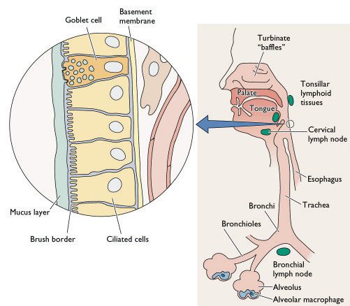
The classical view of measles pathogenesis was that free-virus entered the host through the respiratory route, infecting and primarily replicating within the epithelial cell lining of the respiratory tract. Newly produced virus spreads to nearby lymph nodes where infected monocytes – a type of immune cell - facilitates viral dissemination throughout the host, resulting in the well-known symptoms of measles. The problem with this being that epithelial cells and unstimulated monocytes fail to express the MeV receptor CD150 and infection should therefore not occur. Recently, it has been shown (again using a GFP expressing virus in a macaque model) that MeV predominately infects dendritic cells during the peak of infection, ruling out a major role for monocytes. There is also however no direct evidence of MeV primary replication within the epithelium of the respiratory tract at the early stages of infection. So what exactly happens during the start of infection and does it develop? GFP-expressing viruses may shed light on this question.
[caption id="" align="aligncenter" width="415" caption="Diagramatic representation of the cellular composition of the human respiratory tract - notice the epithelial cell lining and the alveolar macrophages. Dendritic cells are however not shown on this diagram."][/caption]
So how can we study the early stages of infection?
The incubation period of measles is about 2 weeks in humans making it particularly difficult to study the early events of viral infection – the kind of events like host entry, initial site of replication and subsequent intra-host dissemination - this is where we can use a non-human primate model.
Lemon et al generated a highly virulent recombinant MeV based on viral isolates from an outbreak in Sudan; they engineered the viral genome so that it expressed GFP upon entry into cells – an addition that causes little or no replication defects to the virus. Groups of macaques were subsequently infected via the respiratory route allowing highly sensitive visualisation of GFP expressing cells following necropsy. The early time-points of around 5 days post infection were focussed on in this investigation allowing the determination of the early cell targets - epithelium? Immune cells?
So what did they find?
Their results suggest that at the early stages of MeV infection, GFP and hence viral replication is only found in immune cells within the respiratory tract and not the epithelial lining. Dendritic cells and alveolar macrophages are believed to capture viral particles in the lungs allowing spread via infected cells. This is known as a Trojan horse entry mechanism like that used by HIV to pass through mucosal tissues and infect humans - see below. This infection allows for spread and localised replication within nearby lymphoid tissues and then on to draining lymph nodes where massive lymphocyte cell infection may occur facilitating dissemination throughout the host, mainly within lymphoid tissues. Virus can be carried through host blood vessels to other lymphoid target tissues like the tonsils and adenoids and the gut-associated lymphoid tissue ' Peyer's patches'.
[caption id="" align="aligncenter" width="465" caption="HIV entry mechanisms utilising dendritic cells to pass through epithelial cell barriers - the 'Trojan horse' mechanism. This may be directly analogous to MeV entry and primary spread except in the respiratory tract."][/caption]
What does this mean?
This study clearly demonstrates the importance of non-epithelial cells such as dendritic cells in MeV entry, early replication and subsequent systemic spread. It does not however, rule out a major role for epithelial cells in later stages and in transmission - MeV still infects non-CD150 expressing cells and currently the mechanisms of which are unknown. Focusing on the later stages of infection may allow us to appreciate the other cell targets in pathogenesis and viral transmission. As mentioned previously, the use of fluorescent-labeled viruses offers an unprecedented view of viral entry, spread and pathogenic mechanisms. We should look forward to the time when studies like these are applied to other viral and indeed non-viral pathogens.
Lemon, K., de Vries, R., Mesman, A., McQuaid, S., van Amerongen, G., Yüksel, S., Ludlow, M., Rennick, L., Kuiken, T., Rima, B., Geijtenbeek, T., Osterhaus, A., Duprex, W., & de Swart, R. (2011). Early Target Cells of Measles Virus after Aerosol Infection of Non-Human Primates PLoS Pathogens, 7 (1) DOI: 10.1371/journal.ppat.1001263
Coombes, J., & Robey, E. (2010). Dynamic imaging of host–pathogen interactions in vivo Nature Reviews Immunology, 10 (5), 353-364 DOI: 10.1038/nri2746
Through studying viral pathogenesis we seek to understand mechanistically how viral infection and replication causes disease in a particular host. This of course will be subject to a number of complex variables involving both the host and the virus such as: dose; genotype; virus receptor distribution of host tissues;the ability of the virus to replicate in those infected cells and the hosts response to that infection.
Although it is an extremely complex system, knowledge of it may allow us to develop certain preventative strategies alongside new treatments and therapies. That is why being able to study viral pathogenesis is important and why some may welcome a recently published paper reporting initial data (possible cell tropism and host immune response) from a mouse model of infection with xenotropic murine leukemia-related virus (XMRV), a possible novel human pathogen (and a close relation of natural mouse retroviruses).
[caption id="" align="aligncenter" width="258" caption="Mus musculus - relative of Mus pahari. Just incase you forgot what a mouse looked like."][/caption]
Originally identified in a number of human prostate tumour samples, XMRV has since had a conflicting scientific history (covered much better elsewhere), with some studies showing a link between infection and chronic fatigue syndrome (CFS) and prostate cancer. Others since have failed to detect such a link. Despite this, knowledge of how this virus could potentially interact with the human body would of course be useful to acquire.
This understanding has been blocked somewhat by the lack of a small-animal model – a cheap, easier and more ethical alternative to non-human primate studies and of course easier to study than humans. XMRV just does not infect normal lab mice (Mus musculus) and thus pathogenesis in this host does not occur and if pathogenesis doesn’t occur, we can’t study pathogenesis. This species of mouse doesn’t express the receptor that XMRV utilizes to gain entry into cells. Sakuma et al have therefore used Mus pahari, a wild asian relative which does express the receptor for XMRV and thus may tell us something about XMRV pathogenesis.
The group showed that XMRV was able to successfully infect M. pahari cells in vitro and also was able to infect M. pahari following injection of the virus into whole-animals. Following infection, they were able to screen mouse cells and tissues (such as blood) for the telltale signs of XMRV infection over 12 weeks; XMRV being a retrovirus, integrates a DNA copy of its RNA genome into host cells and PCR detection of this integration may allow us to infer XMRV infection. They also investigated the possible role of infectious virus being present in the animal and the of XMRV replication on host cell functioning.
Detection of XMRV DNA in particular tissues may allow us to infer how infection proceeds within the host and how it causes disease. Viral sequences were detected in blood cells, heart, spleen, brain, testis and prostate tissues, although detection was highly variable between mice and a clear picture of infection didn’t really emerge over any of the time periods. The group focused on the effect of XMRV on lymphocytic cell functioning (possibly a good place to start giving its apparent involvement in CFS, an immunological disorder): CD4+ T helper cells and CD19+ B cells being targeted within the spleen. Over all, in some of the mice, increased total white-blood cell numbers was observed early in infection indicating a possible deregulation of lymphocyte development, although this was by no means common and only slight.
The role of both mouse adaptive and innate immunity was also assessed and interestingly, the mice generated a robust antibody response to XMRV antigens yet no long-term studies were involved to see how the virus adapted. Viral genome sequences found within blood and spleen tissues were sequenced and those from the spleen only, displayed predominant G-to-A hypermutation possibly indicative of intra-tissue restriction of viral replication. Host-mediated mutation of viral genomes will therefore more likely result in highly defective viral sequences and possibly prevent future viral infections; one example is that of APOBEC3-like enzymatic reactions.
Of course, these data are rather preliminary with optimisation of infection expected to come later, but at the minute, how does this relate to what happens in humans and will this model actually be on any use?
The development of a permissive small-mammal model of XMRV infection described here will certainly facilitate scientific investigation as it has done for many other viruses although it must be remembered that what happens in a mouse may not happen exactly that way in humans.
This study, however does appear to shed light on cell tropism of XMRV and possibly its transmission - blood-borne (although this was looked at really).
The possible deregulation of host lymphocyte development may play a role in the pathologies associated with it in humans. The highly variable pathological outcomes may be due to the relatively less homogenous gene pool of M. pahari rather than anything to do with the virus - although this may be occurring.
The fact that these mice developed strong antibody responses to this virus may allow the development of vaccine strategies.
But, of course, the importance and relevance of this work will all come down to whether it is or is not a true human pathogen yet we will certainly benefit from this work if it turns out that it is. Expect a lot more from this group if it is.
Sakuma T, Tonne JM, Squillace KA, Ohmine S, Thatava T, Peng KW, Barry MA, & Ikeda Y (2011). Early Events in Retrovirus XMRV Infection of the Wild-Derived Mouse Mus pahari. Journal of virology, 85 (3), 1205-13 PMID: 21084477
Sakuma T, Tonne JM, Squillace KA, Ohmine S, Thatava T, Peng KW, Barry MA, & Ikeda Y (2011). Early Events in Retrovirus XMRV Infection of the Wild-Derived Mouse Mus pahari. Journal of virology, 85 (3), 1205-13 PMID: 21084477





 [/caption]
 [/caption]
 [/caption]
 [/caption]