The mechanisms behind the incredible infectiousness of measles are
poorly understood - that is, until now, where two studies have now come
forward investigating the molecular biology of measles person-person
transmission. Two groups have independently identified the protein, nectin-4 (a cell adhesion molecule) as the receptor allowing measles to infect and emerge from the respiratory tract and spread from person to person, potentially filling in a major gap in our understanding of this important human pathogen.
The measles virus is one of - if not the most - infectious agents currently circulating in human populations. It is also responsible for considerable disease and death worldwide, particularly across the developing world. But luckily, the introduction of a live-attenuated vaccine has significantly reduced it's incidence - this is in spite of a number of recent outbreaks associated with reduced vaccination rates.
There are currently two AIDS pandemics raging across the world: the well-known one in humans and the less recognised one in domestic cats. Feline AIDS, caused by the feline immunodeficiency virus (FIV) - a close relative to HIV - is pretty similar to that afflicting millions of humans and could potentially be used as a model system for studying HIV infection and disease. One major scientific barrier to HIV research in primates is the difficulty in generating transgenic animals efficiently. This, however, could be sidestepped through employing FIV/domestic cat as the experimental system.
The 9,000 year-long relationship between the human species and domestic cats has brought a lot to each respective species: we get rid of nasty pests and enjoy the whole 'cat experience' while they feast on the human-associated rodents and gain some shelter and people love. But what some people might not realise is that the domestic cat has - and will probably continue - to help us in biomedical research. And, for them, the increased research into cat biology may also spill over into non-domestic species, who are on the brink of extinction and might be aided by this work.
Imagine this: Rhinoviruses - one of the culprits responsible for the common cold - enter our body through the upper respiratory tract yet here it stays; the initial and generally the only site of replication is the nasal epithelium. This is how we get a runny/stuffed nose. Contrast this with a virus like nipah virus - a deadly and re-emerging pathogen spread by bats and found across South-east Asia that also enters our body via the upper respiratory tract yet leads to infection and disease in a number of our organs, including the kidneys, blood vessels and the brain, resulting in fatal encephalitis.
How come one virus remains localised while the other goes systemic? And, more specifically how does it transport itself throughout the body?
Well, virus infection of a host is a complex multi-step process involving initial contact and entry into the organism (through the nose) , early replication in particular easy-to-access tissues (upper respiratory tract) and then in some cases the spread to specific tissue sites throughout the body (brain). It is these two stages of replication that most often or not lead to the development of disease yet just how does the virus traverse the gap between the two tissue sites - especially given the minute size of a virus particle?
There are probably three hypotheses of virus spread that could be correct here: lots and lots of virus particles could be released directly into circulation (lymph fluid and blood) from the early site of replication and our blood circulation could do the work for it; the virus could infect those immune cells that cluster around sites of virus infection - or are present naturally in the initial site; or finally, the virus could basically cling on to those highly motile immune cells and be trafficked around the body and effectively transfer infection to the blood vessels and other organs.
Could your white blood cells transport virus throughout your body?
One group has recently asked this question with reference to nipah virus (read the paper here) and has discovered that this virus doesn't infect human immune cells although it does bind to them and this virus/cell interaction facilitates infection of other cells and may allow systemic spread.
They initially came at this problem at an in vitro level - albeit using cells taken directly from the blood of healthy volunteers. The group must have initially thought that nipah must directly infect white blood cells and this is how it spread - after all this is fairly common for other related viruses, such as measles. To determine exactly which cells supported virus replication they added a green fluorescent protein (GFP) - expressing virus to a panel of white blood cells derived from the blood of the healthy humans and specifically assayed for virus-mRNA synthesis, GFP expression and how much virus was released into the culture medium. Surprisingly only one cell type - dendritic cells (DC's) - an antigen-presenting cell - supported any kind of replication and even then it wasn't great (see below).
GFP-nipah virus infects control neuronal U373 cells but not human immune cells - except DC's to an extent
So how come nipah isn't so good at infecting these cells? Is it a receptor issue? Well the group looked at the mRNA levels of the two nipah virus receptors (Ephrin B2 and B3) in all the cells under investigation and found little correlation between their expression and the ability of nipah to infect them. For example, even the dendritic cell which had the lowest level of both receptors is able to support entry and replication while the other cells (macrophages and monocytes) that express higher levels of it fail to do so. The authors hypothesize that the DC's are engulfing nipah virus particles via a process known as macropinocytosis instead of via virus/receptor binding.
I mentioned earlier that the virus doesn't actually need to infect the cells to use them as an effective means of transportation - it can really get by through binding to the outer membrane of the cell much like a microbial hitch-hiker. So they looked at how much nipah virus was associated with each cell following stringent washes and surprisingly all the cells looked at were able to bind nipah virus particles even when they failed to get infected themselves.
But what exactly is blocking infection when the cells bind virus AND express receptor molecules on their surface - something is inhibiting entry. The paper doesn't really address this issue but points to a role of a virus receptor-independent molecule that binds to nipah virus particles yet prevents internalization and engulfment. And even more interestingly, these virus-laden cells were able to efficiently transfer the infectious particles to other cells - as shown with the DC's and PBL's below and this ability to 'trans-infect' was retained over a couple of days (see below).
Transfer of infection with virus bound to immune cells overlaid on top of other cells
OK, so all this work really paints a nice picture of virus infection in the host through the interactions with certain white blood cells that stably bind to - yet fail to get infected themselves - and hence are able to transfer these infectious particles to other cells throughout the body. But this is all cell culture work - no animals have been worked on here so how are we to know if this actually occurs during infection in vivo? Well, the group performed an experiment where-by the mixed nipah with hamster white blood cells and then injected these virus/cells back in to the animals and finally observed whether disease occurred and if so, how bad was it?
Hamster infections with nipah or nipah bound immune cells.
As you can see opposite, by just re-introducing the white blood cells into the animals no death occurred while directly injecting virus into them resulted in 100% mortality but then when the virus/cell mixture was added, these cells were able to transfer the infection to the hamsters with 50% mortality following acute neurological disease reminiscent of that which follows human infection.
So these cell-bound viruses are able to transmit infection in vivo, which points to it having a role in humans.
Now with all this information to hand we can now develop a model as to how nipah infects and causes disease following spread within our bodies - this is known as pathogenesis (see below). Virus could initially enter our body through dendritic cells found within certain epithelium (as the virus could infect these cells in vitro) and then these DC's could traffick to local lymph nodes where virus particles could be released and bind to the white blood cells found there; these cells would then go about their business moving around the body thus transferring infection to a range of different cell types (endothelial cells within blood vessels for example)
Current model for nipah infection and spread within the body
Just one final thought: watch this video below first - imagine this cell going about its normal routine of moving along your blood vessels and squeezing through them but with it covered in infectious virus; every cell ut encounteers will more than likely be exposed to virus and potentially become infected. No wonder nipah is such a deadly pathogen.
Mathieu, C., Pohl, C., Szecsi, J., Trajkovic-Bodennec, S., Devergnas, S., Raoul, H., Cosset, F., Gerlier, D., Wild, T., & Horvat, B. (2011). Nipah Virus Uses Leukocytes for Efficient Dissemination within a Host Journal of Virology, 85 (15), 7863-7871 DOI: 10.1128/JVI.00549-11
Have you ever wanted to visualise viral infection? Ever wanted to observe how they enter and spread throughout their host organism? Ever wanted to know how exactly they caused disease - at the cellular and whole-organism level? Well, this may be entirely possible using fluorescent-labeled recombinant viruses infecting a relevant model system.
Lemon et al recently report the continued investigation of measles virus pathogenesis in a non-human primate (Macaque) model utilising a green-fluorescent protein (GFP) expressing virus. Upon infection of host cells, viral transcription leads to the very high expression of GFP, flooding the cytoplasm with this fluorescent ‘tag’. Subsequent microscopy, imaging and immunohistochemistry allows for the identification and location of the infected cells, tissues and organs - see image above. Tracking of cellular infection allows us to decipher the development of MeV entry, spread and replication at both the cellular and whole-organism level throughout the entire infection. Studies such as these give an unprecedented view of viral infection in a means directed related to that of human infection. This model even allows for macroscopic real-time detection of fluorescence and hence viral infection.
Why is this important for measles?
Despite a highly effective vaccine and significant global control initiatives, measles infection still accounts for significant morbidity and mortality worldwide, mostly in the developing world (164,00 deaths in 2008). This is mostly attributable to the profound immunosuppression induced allowing for further infection with opportunistic pathogens. Currently, much is known about measles pathogenesis yet the molecular mechanisms of such are poorly understood and it is therefore of great interest to better understand these processes by which MeV infects and causes disease in humans. Knowledge of such may facilitate the development of more effective and safer vaccines for measles and indeed other viral pathogens.
Viruses being obligate intra-cellular parasites, must enter and exit cells in order to survive. Most of viral pathogenesis can therefore be attributed to the effects of viral replication of host cells and tissues; a major determinant of which is the expression of receptors on host cells surfaces allowing viral entry, infection and replication. Currently only a single receptor – CD150 - (otherwise known as signalling lymphocyte activation molecule SLAM) has been discovered that wild-type pathogenic MeV uses to enter host cells; the distribution of which only explains part of measles pathogenesis as epithelial and neuronal cells (important target cells) do not express the protein. As indicated by this receptor being expressed on lymphocytes and other immune cells, MeV is a highly lymphotropic virus! But if epithelial cells fail to express the receptor on their surface, how come its possible for MeV to enter via these cells?
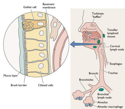
The classical view of measles pathogenesis was that free-virus entered the host through the respiratory route, infecting and primarily replicating within the epithelial cell lining of the respiratory tract. Newly produced virus spreads to nearby lymph nodes where infected monocytes – a type of immune cell - facilitates viral dissemination throughout the host, resulting in the well-known symptoms of measles. The problem with this being that epithelial cells and unstimulated monocytes fail to express the MeV receptor CD150 and infection should therefore not occur. Recently, it has been shown (again using a GFP expressing virus in a macaque model) that MeV predominately infects dendritic cells during the peak of infection, ruling out a major role for monocytes. There is also however no direct evidence of MeV primary replication within the epithelium of the respiratory tract at the early stages of infection. So what exactly happens during the start of infection and does it develop? GFP-expressing viruses may shed light on this question.
[caption id="" align="aligncenter" width="415" caption="Diagramatic representation of the cellular composition of the human respiratory tract - notice the epithelial cell lining and the alveolar macrophages. Dendritic cells are however not shown on this diagram."][/caption]
So how can we study the early stages of infection?
The incubation period of measles is about 2 weeks in humans making it particularly difficult to study the early events of viral infection – the kind of events like host entry, initial site of replication and subsequent intra-host dissemination - this is where we can use a non-human primate model.
Lemon et al generated a highly virulent recombinant MeV based on viral isolates from an outbreak in Sudan; they engineered the viral genome so that it expressed GFP upon entry into cells – an addition that causes little or no replication defects to the virus. Groups of macaques were subsequently infected via the respiratory route allowing highly sensitive visualisation of GFP expressing cells following necropsy. The early time-points of around 5 days post infection were focussed on in this investigation allowing the determination of the early cell targets - epithelium? Immune cells?
So what did they find?
Their results suggest that at the early stages of MeV infection, GFP and hence viral replication is only found in immune cells within the respiratory tract and not the epithelial lining. Dendritic cells and alveolar macrophages are believed to capture viral particles in the lungs allowing spread via infected cells. This is known as a Trojan horse entry mechanism like that used by HIV to pass through mucosal tissues and infect humans - see below. This infection allows for spread and localised replication within nearby lymphoid tissues and then on to draining lymph nodes where massive lymphocyte cell infection may occur facilitating dissemination throughout the host, mainly within lymphoid tissues. Virus can be carried through host blood vessels to other lymphoid target tissues like the tonsils and adenoids and the gut-associated lymphoid tissue ' Peyer's patches'.
[caption id="" align="aligncenter" width="465" caption="HIV entry mechanisms utilising dendritic cells to pass through epithelial cell barriers - the 'Trojan horse' mechanism. This may be directly analogous to MeV entry and primary spread except in the respiratory tract."][/caption]
What does this mean?
This study clearly demonstrates the importance of non-epithelial cells such as dendritic cells in MeV entry, early replication and subsequent systemic spread. It does not however, rule out a major role for epithelial cells in later stages and in transmission - MeV still infects non-CD150 expressing cells and currently the mechanisms of which are unknown. Focusing on the later stages of infection may allow us to appreciate the other cell targets in pathogenesis and viral transmission. As mentioned previously, the use of fluorescent-labeled viruses offers an unprecedented view of viral entry, spread and pathogenic mechanisms. We should look forward to the time when studies like these are applied to other viral and indeed non-viral pathogens.
Lemon, K., de Vries, R., Mesman, A., McQuaid, S., van Amerongen, G., Yüksel, S., Ludlow, M., Rennick, L., Kuiken, T., Rima, B., Geijtenbeek, T., Osterhaus, A., Duprex, W., & de Swart, R. (2011). Early Target Cells of Measles Virus after Aerosol Infection of Non-Human Primates PLoS Pathogens, 7 (1) DOI: 10.1371/journal.ppat.1001263
Coombes, J., & Robey, E. (2010). Dynamic imaging of host–pathogen interactions in vivo Nature Reviews Immunology, 10 (5), 353-364 DOI: 10.1038/nri2746


 [/caption]
 [/caption]
 [/caption]