Field of Science

A Mouse Model of XMRV Pathogenesis?

Through studying viral pathogenesis we seek to understand mechanistically how viral infection and replication causes disease in a particular host. This of course will be subject to a number of complex variables involving both the host and the virus such as: dose; genotype; virus receptor distribution of host tissues;the ability of the virus to replicate in those infected cells and the hosts response to that infection.

Although it is an extremely complex system, knowledge of it may allow us to develop certain preventative strategies alongside new treatments and therapies. That is why being able to study viral pathogenesis is important and why some may welcome a recently published paper reporting initial data (possible cell tropism and host immune response) from a mouse model of infection with xenotropic murine leukemia-related virus (XMRV), a possible novel human pathogen (and a close relation of natural mouse retroviruses).

[caption id="" align="aligncenter" width="258" caption="Mus musculus - relative of Mus pahari. Just incase you forgot what a mouse looked like."][/caption]

Originally identified in a number of human prostate tumour samples, XMRV has since had a conflicting scientific history (covered much better elsewhere), with some studies showing a link between infection and chronic fatigue syndrome (CFS) and prostate cancer. Others since have failed to detect such a link. Despite this, knowledge of how this virus could potentially interact with the human body would of course be useful to acquire.

This understanding has been blocked somewhat by the lack of a small-animal model – a cheap, easier and more ethical alternative to non-human primate studies and of course easier to study than humans. XMRV just does not infect normal lab mice (Mus musculus) and thus pathogenesis in this host does not occur and if pathogenesis doesn’t occur, we can’t study pathogenesis. This species of mouse doesn’t express the receptor that XMRV utilizes to gain entry into cells. Sakuma et al have therefore used Mus pahari, a wild asian relative which does express the receptor for XMRV and thus may tell us something about XMRV pathogenesis.

The group showed that XMRV was able to successfully infect M. pahari cells in vitro and also was able to infect M. pahari following injection of the virus into whole-animals. Following infection, they were able to screen mouse cells and tissues (such as blood) for the telltale signs of XMRV infection over 12 weeks; XMRV being a retrovirus, integrates a DNA copy of its RNA genome into host cells and PCR detection of this integration may allow us to infer XMRV infection. They also investigated the possible role of infectious virus being present in the animal and the of XMRV replication on host cell functioning.


Detection of XMRV DNA in particular tissues may allow us to infer how infection proceeds within the host and how it causes disease. Viral sequences were detected in blood cells, heart, spleen, brain, testis and prostate tissues, although detection was highly variable between mice and a clear picture of infection didn’t really emerge over any of the time periods. The group focused on the effect of XMRV on lymphocytic cell functioning (possibly a good place to start giving its apparent involvement in CFS, an immunological disorder): CD4+ T helper cells and CD19+ B cells being targeted within the spleen. Over all, in some of the mice, increased total white-blood cell numbers was observed early in infection indicating a possible deregulation of lymphocyte development, although this was by no means common and only slight.

The role of both mouse adaptive and innate immunity was also assessed and interestingly, the mice generated a robust antibody response to XMRV antigens yet no long-term studies were involved to see how the virus adapted. Viral genome sequences found within blood and spleen tissues were sequenced and those from the spleen only, displayed predominant G-to-A hypermutation possibly indicative of intra-tissue restriction of viral replication. Host-mediated mutation of viral genomes will therefore more likely result in highly defective viral sequences and possibly prevent future viral infections; one example is that of APOBEC3-like enzymatic reactions.

Of course, these data are rather preliminary with optimisation of infection expected to come later, but at the minute, how does this relate to what happens in humans and will this model actually be on any use?

  • The development of a permissive small-mammal model of XMRV infection described here will certainly facilitate scientific investigation as it has done for many other viruses although it must be remembered that what happens in a mouse may not happen exactly that way in humans.

  • This study, however does appear to shed light on cell tropism of XMRV and possibly its transmission - blood-borne (although this was looked at really).

  • The possible deregulation of host lymphocyte development may play a role in the pathologies associated with it in humans. The highly variable pathological outcomes may be due to the relatively less homogenous gene pool of M. pahari rather than anything to do with the virus - although this may be occurring.

  • The fact that these mice developed strong antibody responses to this virus may allow the development of vaccine strategies.


But, of course, the importance and relevance of this work will all come down to whether it is or is not a true human pathogen yet we will certainly benefit from this work if it turns out that it is. Expect a lot more from this group if it is.

ResearchBlogging.org

Sakuma T, Tonne JM, Squillace KA, Ohmine S, Thatava T, Peng KW, Barry MA, & Ikeda Y (2011). Early Events in Retrovirus XMRV Infection of the Wild-Derived Mouse Mus pahari. Journal of virology, 85 (3), 1205-13 PMID: 21084477

Sakuma T, Tonne JM, Squillace KA, Ohmine S, Thatava T, Peng KW, Barry MA, & Ikeda Y (2011). Early Events in Retrovirus XMRV Infection of the Wild-Derived Mouse Mus pahari. Journal of virology, 85 (3), 1205-13 PMID: 21084477

Seeing the big picture of RNA virus evolution

This post was chosen as an Editor's Selection for ResearchBlogging.org

From both a medical and a scientific viewpoint, the evolution of viruses is extremely important to us;  viral adaptation to their ever changing environment is responsible for major morbidity and mortality worldwide so maybe studying this  may allow us to predict virus evolution in the future and may help prevent pandemics occuring?

We kind of know a lot about how viruses evolve at the small-scale; we know how viruses generally create genetic diversity - mutations - and how processes such as natural selection and genetic drift act on these genetic changes and influence the way viral genomes change over time. What we don't know however, is how viruses change at the larger-scale - how these above processes influence viral genomes over thousands of years, including: how and why viruses speciate, how their genome structure evolves and how and when do new viruses originate.

[caption id="" align="aligncenter" width="375" caption="An example phylogenetic tree - The paramyxoviridae containing a number of important human and animal pathogens. Notice the host/viral species distribution."][/caption]

A recent study, investigating the evolution of a number of RNA viruses has sought to reconcile this lack of understanding by attempting to assess virus 'macroevolution'- specifically viral speciation. By generating large and highly robust phylogenetic trees (using significantly highly conserved amino-acid sequences of a single viral protein) for 5 genera of RNA virus including: the Alphaviruses, Caliciviruses, Paramyxoviruses, Rhabdoviruses and the Flaviviruses, the team were able to map the host species of each virus species onto the trees and this allowed them to infer the mode of speciation of each virus genus.

More specifically they asked: Do closely related viral species infect the same host and are therefore believed to have speciated in that host or do they infect completely different hosts which are believed to have speciated following host jumping?

What is a virus species?


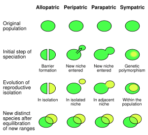
[caption id="" align="aligncenter" width="300" caption="General modes of speciation - we may think of viruses speciating by either allopatric (host shift) or sympatric (intra-host divergence)."][/caption]

The concept of the viral species has been a hard one to determine becuase viruses don't reproduce sexually. It is generally thought to be rather a arbitrary classification, however, most virus 'species' tend to be phylogenetically and often phenotypically stable genetic lineages and hence may be thought of as 'biological relevant'. We may think of viral speciation much like we think of speciation in the classic sense: allopatric or 'geographical speciation' (virus adaptation to a new host species) and sympatric - that not requiring the forces of georgaphic isolation (generation of viral speciation within a single host). Virus sympatric speciation requires the adapation to a new infectious niche within a host, for example a new lineage may infect new cell types within that host. Virus allopatric speciation requires host-jumping or adaptation to a new host altogether but may result from co-divergence follwoing host speciation. Both processes may result in two or more 'stable, phylogenetic and phenotypic genetic lineages. But what does the data say about it - how do viruses evolve in the real world?

What the data says


The results were split - at leats 50% were found to have 'speciated' via sympatric-like processes and half from allopatric-like processes. The group stress, however, that a major caveat of this study is that it highlights our limited understanding of what specific host speces particular viruses infect; in this study most hosts were classified as 'birds' or 'plants' or 'Carnivores' which limits the resolution of phylogenetic studies and leads to the overestimation of sympatric speciation events which would otherwise not exist if exact hosts were known. This leads us to put little confidence on our earlier 50/50 estimate and most likely the role of sympatric speciation would be a lot less important than allopatric modes of speciation in reality.

Why do RNA viruses evolve this way - What controls viral speciation?


So, we may say that most RNA virus speciation is caused by allopatric modes - or host jumping, but this may seem counterintuitive as there are some major barriers to viral emergence. The group argue, however, that it may take a lot more - genetically speaking - for a virus to speciate within a host than it does for a virus to jump species - eg. replicate in a new cell type/alter antigenic epitopes. The apparant preference for allopatric speciation may be controlled by intrinsic biological factors of these RNA viruses, namely: their extremely small genome size which effectively constrains evolutionary innovation. Those changes required for host jumping (change in receptor binding sites for instance) may be relatively minor when compared to those and the more closely related the host species are then the more easily host-jumping will occur - which is what we see here.

This study highlights the key role that viral 'allopatric' speciation or host - jumping plays in the evolution of RNA viruses yet further emphasizes the need to better study and understand viral biodiversity and host range in the wild - not only focusing on those medically important human viruses. Further research may be carried out on the molecular barriers to both cellular and host switching for these RNA viruses. This study will act as a model system that may be applied to other viral lineages - what about the RNA viruses with segmented genomes? What about the DNA viruses? Retroviruses?

ResearchBlogging.org
Kitchen A, Shackelton LA, & Holmes EC (2011). Family level phylogenies reveal modes of macroevolution in RNA viruses. Proceedings of the National Academy of Sciences of the United States of America, 108 (1), 238-43 PMID: 21173251

Measles, Papua New Guinea and the brain

This post was chosen as an Editor's Selection for ResearchBlogging.orgYou may not have realised that - since most people nowadays have been vaccinated against it and have never seen it - but measles is a very serious illness. Generally an acute disease of children, measles is spread by the measles virus where it infects the body via the respiratory route and establishes a systemic infection - involving multiple organ systems - via your bodies own immune cells leading to the typical rash, mild to severe respiratory distress and immunosuppression (Rima and Duprex 2006).

[caption id="" align="aligncenter" width="463" caption="Measles virus replicative cycle"][/caption]

In the 'developed' world we tend not to think about infectious disease in the same way as people in other parts of the world; national vaccination campaigns have largely removed the threat (not considering some minor outbreaks) of the some of the biggest human killers and we no longer worry ourselves over whether a family member will come down with these diseases.

Subacute Sclerosing Panencephalitis or SSPE is one of the most serious complications of measles resulting from viral infection of the central nervous system; SSPE is rare (1 in 10,000-25,000 measles infections) but is almost always fatal. Following infection at a particularly young age and on average 8 years following acute infection, a progressive deterioration of neurological function presents : loss of attention span, uncontrolled movements, behavioural changes, cognitive impairment and in all cases vegetative state is entered and death occurs.

It is caused by persistent measles infection i.e one that the isn't removed when your immune system kicks in, which spreads throughout the  cells found within the brain causing cell death and inflammation. Strangely, no infectious virus can be recovered from infected brains and when this was investigated further they found that many mutations occurred throughout the genome rendering many of the genes nonfunctional. Although the major replicative functions (replication and gene expression) were left intact, the genes required for normal particles formation were those mutated suggesting that the virus may exploit the unique cellular environment in the CNS to spread, replicate and survive.

[caption id="" align="aligncenter" width="402" caption="Green Fluorescent Protein expressing measles virus infection of neuronal cell"][/caption]

As I mentioned previously, due to increased transmission of virus, poverty and poor nutrition, measles infection is extremely serious in developing countries and it is no surprise that SSPE occurs here in higher numbers. In Papua New Guinea there exists a very high incidence of SSPE, THE highest incidence - roughly 3 - 20 times as many cases are reported (98 per million people versus 5 per million people). Manning et al (2011) have attempted to further characterise SSPE behaviour in this country between 1997 and 2008 and highlights the significant burden that measles is in many developing countries. They measured SSPE incidence, measles infection rates and time of birth of each patient presenting with SSPE finding a direct correlation between time of birth, measles epidemics and presenting with SSPE. The group emphasises the requirement

Why is SSPE incidence so high here and what can we do about it? SSPE rates are linked to measles infections in a population and hence have been significantly reduced following measles vaccination campaigns. Sadly, only half of children in Papua New Guinea receive two measles vaccines prior to 1st birthday - not enough to sufficiently protect an individual nor a population from measles infection and hence SSPE; there is insufficiently low-level of herd immunity in regions such as papua New Guinea. The level of vaccine effectiveness of measles vaccine in this region is also particularly low - possibly reflecting damage to the vaccine from cold-chain disruption (in tropical climates it is difficult to keep vaccines refrigerated), population genetic effects or persistence of low-level non-neutralising maternal antibody.

We can no longer afford to ignore the importance of measles in developing countries like Papua New Guinea and we must stress the need for adequate vaccine effectiveness and coverage in already susceptible human populations. Studies like these with SSPE emphasise the real-world need for the investigation of the molecular mechanisms of measles virus persistence and we should look forward to a time when we can adequatly treat measles CNS complications - or maybe with better vaccination coverage we may not have to worry about this.

Manning, L., Laman, M., Edoni, H., Mueller, I., Karunajeewa, H., Smith, D., Hwaiwhanje, I., Siba, P., & Davis, T. (2011). Subacute Sclerosing Panencephalitis in Papua New Guinean Children: The Cost of Continuing Inadequate Measles Vaccine Coverage PLoS Neglected Tropical Diseases, 5 (1) DOI: 10.1371/journal.pntd.0000932

Rima, B., & Duprex, W. (2006). Morbilliviruses and human disease The Journal of Pathology, 208 (2), 199-214 DOI: 10.1002/path.1873

Viral nanotechnology - at the virus-chemistry interface

Viruses cause death and disease - Avian Influenza, Swine-origin Influenza, HIV, HPV, measles..... its hard to imagine viruses doing anything else - right?

But viruses don't have to cause disease - they can infect, replicate and exit without the host even realising it was there. Another view of viral infection is that we can exploit this very nature of viruses for our own means - meet: viral engineering (one flavour of biologically inspired nanotechnology).

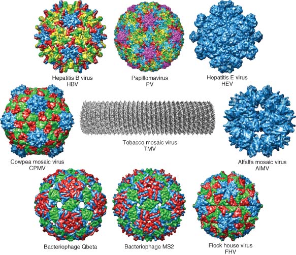
[caption id="" align="aligncenter" width="352" caption="Viral nanoparticles: the diversity"][/caption]

Viruses are basically self-assembling storage containers that can enter and exit cells and deliver their contents, they are very small, are biodegradable, can be modified (relatively) easily and have an excellent ability to travel around the human body - one big bonus is that in some cases (plant viruses) they are also extremely cheap.

A recent review describes these 'viral-nanoparticles' (VNPs) as:
....dynamic, self-assembling systems that form highly symmetrical, polyvalent, and monodisperse structures. They are exceptionally robust, they can be produced in large quantities in short time, and they present programmable scaffolds. VNPs offer advantages over synthetic nanomaterials, primarily because they are biocompatible and biodegradable. VNPs derived from plant viruses and bacteriophages are particularly advantageous, because they are less likely to be pathogenic in humans and therefore less likely to induce undesirable side effects.

Of course there are many caveats with these applications such as we would have to thoroughly test the toxicity (including cell death and immunogenicity) of such VNPs as human pathogens may have been used as the basis of the design, although the use of plant viruses may circumvent these dangers. The pharmacokinetics, infectivity and replication of viruses will be assessed in animal models prior to use as so will the stability in both a physical and genetic sense. Yet there are plenty of uses for VNPs that would not have to be anywhere near a human patient.

Despite these difficulties, we have a great chance of developing improved VNPs through the application of genetic engineering and chemical modifications, allowing us to generate novel combinations of genes and properties into a single viral particle. We no longer have to rely on 'wild-type' virus genomes - we can improve on what is out there. By applying a better understanding of natural viral pathogenesis including cell entry, replication, gene expression, cellular tropism and immunomodulation we should be able to rationally design safer, more efficacious and cheaper VNPs for whatever purpose we want. We can now begin to think of viruses as a novel materal that can altered to generate improved properties and thinking this way should open up many possibilities for medicine, industry and science. This is a basic tenet of synthetic biology.

Synthetic biology meet virology.


As of today, this research has been moving at an extremely fast pace - viruses are now used in cancer treatments, bacteriophages have been used to kill off bacterial infections, viruses have been applied in materials science, improved electronics have been developed using viral particles and targeted viruses have been used in biomedical imaging technology. Yet as our understanding of virus/host interactions increases and research on the applications of these VNPs begins to move from in vitro to in vivo investigations we will see more and more uses for these novel materials in both the clinic and in industry. Look forward to the future of viral nanotechnology!

As the review finishes off:
The virus-chemistry interface remains an exciting place to be!

N.F. Steinmetz, Viral nanoparticles as platforms for next-generation therapeutics and imaging devices. Nanomedicine: NBM 2010;6:634-641, doi:10.1016/j.nano.2010.04.005

Infectious disease meets the epigenome

The study of epigenetics (see Pharyngula's excellent article) has allowed us to see biology and genetics through new eyes. The fact that heritable traits can be encoded in not only the nucleic acid sequences of As, Cs, Ts and Gs but also in the physical conformation of chromosomes and chemical modification of DNA has added a new level of complexity to our understanding of life. Covalent modifications to both DNA and associated histones and chromatin can result in the formation of active or repressed genetic regions; transcription of these genes found in that area is thus activated or repressed. Embryonic development, behaviour and cancer formation  have all been impacted by the discovery of this new genetic system wit deregulated epigentic processes leading to the development of these diseases - but what about in infection, immunity and pathogenesis of associated diseases?

[caption id="" align="aligncenter" width="300" caption="Epigenetics: DNA, histones, ovalent modification and chromatin."][/caption]

Uncovering the biomedical and evolutionary importance of primate innate immunity


Primates such as chimpanzees differ in the diseases that affect them - especially when we compare them to those that affect us. Progression to AIDs, cancer incidence, Alzheimers disease and malaria either do not affect or cause less severe diseases in non-human primates than they do in humans. So why are our primate cousins so differently affected by these particular diseases than we are? What causes these differences? Are they environmentally mediated? Behavioural? Molecular?

What does the innate immune system have to say about it?

The pathogenesis of disease is an important area of research when trying to understand the origins and progression of diseases. It depends on many variables - host specific, environmental or pathogen specific. A recent publication has sought to understand these differences from the point of view of host factors mainly the innate immune system. By comparing genome-wide gene expression patterns in primary immune cells (monocytes) cultured in vitro from groups of humans, chimpanzees and rhesus macaques and applying bioinformatic analysis to the results, the investigators were able to detect those genes whose expression is altered upon stimulation with lipopolysacharide (LPS), an important activator of the innate immune system. From this data set they could ask, functionally, what makes the human immune system different?


3,170 genes were seen to be differentially expressed with 793 changing in all three species indicating a conserved function. Other genes showed species-specific changes allowing researchers to ask what makes each species unique? More importantly what makes non-human primates different when it comes to diseases? There were 335 genes in the human monocytes that were expressed differently and these were divided between particular pathways.

What does this data have to say about specific diseases?

Those genes listed as being involved in viral infection were those most likely to be species-specific indicating the rapid adaptation of host immunity to fast-evolving viral pathogens. The data sheds light on the possible lo incidence of cancer in non-human primates by detecting the difference in apoptosis/cancer related genes - although the signifigance of monocyte gene expression when considering the whole-organism in diseases such as cancer is difficult to say. Most interestingly seen are those genes involved in HIV infection and AIDs  whose expression may explain why chimpanzees do not progress to AIDs or do so slowly.

Despite the problems in culturing non-human primate immune cells in vitro and the difficulties in controlling for environmental effects (different diets of primates), this study goes some way to understanding at a functional level what makes humans human from an immunological point of view. Although focussing on a single cell type, the monocyte, other cells may prove useful in investigating innate differences - as other cells also function in innate immunity - mucosal epithelium for one. This work paves the way for more detailed molecular analysis but also of more genome-wide work looking at other cells, other activators and pathogens (not LPS but HIV?). Understanding what makes humans different in a pathogenic light should focus not just on immunology at the gene expression level but also differences in epigenomics, behaviour, anatomy and cell biology.


Varki, Aijit. 2000. A Chimpanzee Genome Project is a Biomedical Imperitive. Genome Res. 2000. 10: 1065-1070. doi: 10.1101/gr.10.8.1065

Barreiro, Luis B., John C. Marioni, Ran Blekhman, Matthew Stephens, and Yoav Gilad. 2010. Functional Comparison of Innate Immune Signaling Pathways in Primates. Ed. Greg Gibson PLoS Genetics 6, no. 12 (December): e1001249. doi:10.1371/journal.pgen.1001249. http://dx.plos.org/10.1371/journal.pgen.1001249.