Field of Science

On the experimental generation of endogenous (non-retroviral) RNA viruses

A retrovirus. http://www.itqb.unl.pt/
The sheer amount of genomic data now available from a wide range of species has allowed the increased scrutiny over what genes and DNA sequences are present in their chromosomes. What we have begun to notice is that many of these sequences have a viral origin. 

And, in the recent half-decade, the numbers of these endogenous viruses discovered have rapidly increased, but how did they get there? What are they doing? And, are they bad for us? Only a true experimental model system can answer these question but this is something which is lacking.


Lets talk about ERVs

Now, viruses have left their mark on our genomes in more ways than one; infection and associated disease/mortality has heavily influenced the genetic structure of populations via natural selection and genetic drift for millions of years. Yet, another important mechanism is that employed by the endogenous retroviruses (ERVs) that have inserted a DNA copy of themselves into our chromosomes - the norm for retroviruses - and have forever become part of us.


Over the course of evolution, these once infectious viruses have become redundant, building up a collection of genetic mutations resulting in loss of replicative ability. Although many still play a role in the cellular biology of the host and have been a great source of genetic novelty over the billions of years of evolution.


For some excellent info on these viruses, see ERVs archive of ERV-related material.


What about non-ERVs?

However, what we have noticed is that viruses other than retroviral species have inserted themselves into genomes of humans, other animals and even plants and fungi. Many of these viruses have a DNA phase in their replication cycle, which is put into the genome of their host to aid their survival and so it may not be all that surprising that they have stayed with us through evolution (these viruses include many single-stranded DNA viruses and again).

One intriguing observation is that many of these non-retroviral endogenous viruses are in fact - or were - RNA viruses with no known DNA phase during replication. They are therefore called Non-retroviral RNA virus sequences (NRVSs). See plant NRVSs and mammalian NRVS (ebola virus-like , borna virus-like  and many more - (lots, they're everywhere). There is strong evidence that these integrations occurred thousands, if not millions of years ago and could have played a role in the evolution of many species.


How can we study these viruses?

But just exactly how do these viruses do it? After-all, they are RNA viruses without a reverse-transcriptase enzyme and hence no natural ability to produce a DNA genome that can be inserted into our chromosomes. And, can we follow this endogenisation experimentally? One mechanism is thought to occur when an endogenous retrovirus-like element joins itself to a non-endogenous RNA virus and then this chimera is put into our genome. But this is really only half the story - can we ever study the entire process, from initial infection to endogenisation?

 For an RNA virus to become fully integrated into our germline it has to first infect our germ-line cells (sperm/oocytes); its RNA genome must be copied into DNA and this DNA molecule must be inserted into the chromosome. It also must allow for the development of healthy and reproductively active offspring and can then let evolution take its course. An experimental model system of this process would allow for a better understanding of this process in molecular detail and how this relates to the evolutionary process as a whole.

Here's how you would do it:

The animal model

Bank vole - a good model for endogenous viruses?
A small-animal model that could be infected by a  type of virus that had been shown to integrate into the genome (borna disease virus, for example) would make this easier to study. Plus, many rodents have been shown to harbour many NRVSs already.

The virus infection

You would infect the animals with the virus in as natural conditions as possible and look to see whether the virus entered and replicated in the cells of the germ-line.A GFP-expressing virus would work best for this.

Detection of RNA - DNA

What you would have to do is be able to track the process of turning the RNA genome into DNA. A PCR-based screening would work well for this and could be applied to a range of tissues in the host, including occytes/spermatozoa.

Integration

To prove that the DNA copy was inserted into the host chromosome you would need to sequence the sites where the DNA had integrated in and determine where in the genome it lay.

Stability

This experimentally infected rodents could be bred continously and the presence of endogenised virus looked for in their offspring. The expression of said virus genes (if there is any) could be followed in rodent tissues.

Borna disease PCR without reverse-transcriptase. A) no nuclease treated, B) RNA nuclease treated, C) DNA nuclease treated and D) PCR with reverse transcriptase step

Well one paper has maybe taken the first step in the development of such a model system (although they may not know it). It has shown evidence that if you infect baby bank voles with borna virus, directly into their brain you can detect borna virus-specific DNA sequences using PCR following DNA extraction (see above PCR gel for results).  And, these sequences resulted from the virus, not some already-endogenised borna virus sequence. Although they did not check for germ-line infection or integration, this is the first step. The applicability of Borna virus reverse genetics and these animal models could make this kind of study feasible but certainly not easy. We may in future catch a glimpse of this process in real-time.

ResearchBlogging.orgKinnunen, P., Inkeroinen, H., Ilander, M., Kallio, E., Heikkilä, H., Koskela, E., Mappes, T., Palva, A., Vaheri, A., Kipar, A., & Vapalahti, O. (2011). Intracerebral Borna Disease Virus Infection of Bank Voles Leading to Peripheral Spread and Reverse Transcription of Viral RNA PLoS ONE, 6 (8) DOI: 10.1371/journal.pone.0023622

How come direct cell-cell spread of HIV allows ongoing replication in the face of antiviral therapy?

Our struggle with HIV/AIDS epitomizes societies' millennia-old fight with microbial pathogens. One goal of HIV research is to generate effective interventions that will allow us to: 1) prevent further spread of HIV (vaccines and behavioural changes), and 2) eliminate the  virus from those already infected (antivirals).

This sterilizing immunity - as it is referred to - has been a long sought-after goal for a number of viruses, yet widespread use of highly-active anti-retroviral therapy (HAART)  fails to completely remove the virus from an individual patient. Somewhere, somehow HIV is continuously replicating in your body. But how and why is this possible?

The spread of HIV from Dendritic cells to T lymphocytes or T to T cells may allow escape from antivirals. http://pathmicro.med.sc.edu

One potential mechanism is that HIV lies in a latent state - anatomical (central nervous system) or biochemical (following DNA integration and before gene expression) - where these drugs cannot inactivate it.

But researchers, headed by David Baltimore at the California Institute of Technology, have come forward with both theoretical and experimental evidence suggesting a novel mechanism that explains how the virus may be able to circumvent HAART treatment through continuous replication and direct cell-cell spread. Their results were published in Nature last week. See here.

What is cell-cell spread?

Viruses can infect new cells via a number of mechanisms. The most well-characteristic being via cell-free virus particles (see computer simulation paper here). In this, new viruses are released from the originally infected cell and diffuse to an uninfected one nearby, thus establishing a novel infection.The problem with this is that it is pretty inefficient. For one, the viruses could be carried anywhere and even if they reach a cell, it may not be the right one. One other, more efficient means of transmission is through direct cell-cell spread, which generally takes diffusion out of the picture.
Model of how HIV moves from cell-cell. A) Dendritic cell (DC) with natural fold-like projections. B) DC picks up virus particles in red C) virus is held in vesicles within cell, D) T cell projections induced, E) formation of the virological synapse, F) release, binding and entry of HIV into T cells. (Felts, et al 2010)

HIV and its close relative, the human T lymphotropic virus (HTLV-1) have been shown to move from one cell to another through this mechanism.

Original paper here

Initial visualisation

In-depth 3D structural analysis

Check out the great videos here


Following interaction with dendritic cells or T cells, the virus directs the assembly of a structure (referred to as the virological synapse) linking the two cells via alterations of the cytoskeleton which causes the two plasma membranes to come close together. This structure initially derives from the target cell.

It is here where new virus particles are released into the gap that has formed, thereby increasing the efficiency of spread by directing transmission and limiting the effects of diffusion.This synaptic structure, ultimately acts to concentrate both virus and receptors at defined subcellular locations. This kind of spread may allow the virus to become essentially invisible to our bodies' immune defences, such as neutralizing antibodies, and even antiviral drugs.

How does this allow escape from treatment?

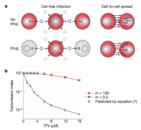
Their model: a) inefficient cell-free infection will be eradicated with antvirals. The more efficient direct cell-cell spread will persist as it involves many more virus particles. b) The mathematical model (backed up by experimental data) showing the loss of infection (transmission index) with increasing concentration of antiviral (TFV) when we have few (m =0.2) or many (m = 100) virus particles.
Expanding the model

Initially, they generated - and experimentally verified - a mathematical model of how HIV transmission may differ during antiretroviral treatment under cell-free (inefficient - low virus transmission) or direct cell-cell spread (efficient - high virus transmission) mechanisms. Their results indicate that when there are many viruses around, the infection is more resistant to the drugs. This, they suggest, is due to the increased probability that at least one virus particle will not interact with the antiviral.


This observation extended to direct cell-cell spread when they introduced previously infected cells with non-infected ones and measured transmission of virus. Both the experimental and theoretical modelling suggest that this spread could be responsible for the ongoing replication seen in patients being treated with antivirals.


What does this mean for HIV therapy? Well, this work brings experimental evidence on the already known process of ongoing HIV replication in the face of antivirals. Through experimentally identifying this mechanism, the researchers here have uncovered a weak-point in HIV biology, one that is not currently being targeted. This method of cell-cell spread may also facilitate escape from neutralizing antibody responses through physically preventing their interaction with HIV proteins. How then might our own bodies combat this virus if its ability to more from cell-cell was inhibited? This work further adds to the growing body of work highlighting direct cell-cell spread as a principle mode of transmission for these retroviruses.

ResearchBlogging.orgSigal A, Kim JT, Balazs AB, Dekel E, Mayo A, Milo R, & Baltimore D (2011). Cell-to-cell spread of HIV permits ongoing replication despite antiretroviral therapy. Nature PMID: 21849975

When you say flu, what do you really mean?

Everyone knows the 'flu when they see it. Its like the cold but only worse, right? But often the public are very quick to brand something the flu when it clearly isn't and this even extends to doctors in the clinic. As highlighted by this recent paper, published in BMC Infectious Diseases.

BioMed Central | Full text | During the summer 2009 outbreak of "swine flu" in Scotland what respiratory pathogens were diagnosed as H1N1/2009?

Turns out a lot of viral infections look like influenza, clinically. But what are doctors misdiagnosing as flu? Based on this study, which paired molecular diagnostics with what the clinic thought each patient had  based on well-defined guidelines (mostly flu at the early stages of the 2009 swine-influenza pandemic - April - July).


Are these symptoms clinically specific to influenza infections? http://symptomswineflu.com 


What they did was:


We examined the results from 3247 samples which were sent to the laboratory during April-July 2009
And,
Real-time RT-PCR was carried out in order to detect influenza A (a generic assay and a H1N1/2009 specific assay [7]), B and C, RSV, rhinovirus, parainfluenza 1-4, human metapneumovirus, coronavirus (229E, NL63, HKU1 and OC43), adenovirus, and Mycoplasma pneumoniae.
 Out of the 3247 samples analysed, 27.9% were diagnosed as being infected with a respiratory pathogen they looked for, what is causing their symptoms??. While human rhinovirus infections were the most common, H1N1 influenza infection was one of the least common at 0.8%. A whole heft of viral and bacterial pathogens were detected at levels intermediate to these.


So, whats going on here? How come it's so difficult to diagnose these guys and after-all, does it really matter?


Well, this work highlights the difficulty in accurately determining the causative pathogen from basic symptoms. The viruses looked at in this study all share replicative and epidemiological characteristics: respiratory pathogens, infect the upper/lower respiratory tract and, as viruses, induce a general interferon/inflammatory response (sore throat, headache, malaise, fever, muscle pains etc.) and are transmitted among the winter/spring months. It is not until you look at the molecular level, that you realise they are all at least slightly different.


And, yes it does really matter. Many of these people may have developed life-threatening symptoms as a consequence of an infection that was not influenza and would have been missed by the clinicians. Also, at a great cost to society and the patient (financial, medical and emotiona;), some people were wrongly treated with antiviral medication after being diagnosed swine flu.


What studies like this show, is the importance of molecular diagnostics in times of pandemic infections. But the problem here is that, despite their high specificity, they can be expensive and difficult to run, especially in 'field' locations across the world. 

miRNAs, viruses and high blood pressure

You may have read earlier this week how human cytomegalovirus - a seriously common pathogen - was discovered to be a possible cause of high blood pressure. What you may not have heard about (or read), is the actual paper that these headlines refer to - see paper here. And, if you had, you may not have been so quick to jump to that same conclusion the media had for it was only a correlation - no causation was determined - but the results are nonetheless important, as you'll see below.

Human cytomegalovirus infected endothelial cell with various viral proteins fluorescently labelled. Does HCMV cause hypertension? http://www.princeton.edu/artofscience. By Joerg Schroeer

What is hypertension?

Hypertension - or high blood pressure - is a chronic disease with deadly implications, affecting an estimated one billion adults worldwide. Referred to as a global pandemic, if left unchecked, high blood pressure can result in terminal damage to both blood vessels and organs leading to stroke, heart attack or kidney failure. And, as if this weren't bad enough, it kills silently while if detected early enough it can be effectively controlled through various drug regimes and lifestyle changes.

 Two flavors of hypertension are currently recognized: essential - the most common sort (90%) and secondary, the more rarer form. The causes of secondary are well characterized and usually result from a number of acute illnesses, while the origins of essential are more difficult to determine. This most likely reflects the complex nature of the disease, which results from interactions from both genetic and environmental components; any one of these in isolation having little effect on its overall state.

Given its global importance, the factors predisposing to - or causing - essential hypertension are subject to intense research, making the identification of a viral origin of the disease all the more interesting.

What about the virus?

Typical herpesvirus (of which HCMV is a member) particle morphology. http://viralzone.expasy.org


HCMV, a member of the herpesvirus family of viruses (large, double-stranded DNA viruses with over one hundred genes) is an expert human pathogen. And, with 40% percent of the human population estimated to be infected with the virus, who can argue. Also based on seroprevalence, 80% of the elderly population are infected making it one serious human pathogen.

Following initial infection, HCMV has the ability to rapidly enter a latent state, persisting within specific cells inside your body. The usual cells are fibroblasts, endothelial cells and some white blood cells. From here, it will every-so-often re-activate itself and be shed in your saliva, ready to infect the next susceptible person. So once you get infected, you are always infected. Although it barely causes disease in those with fully functioning immune systems, the people who are immuno-compromised (neonates, AIDS sufferers, chemotherpy patients or the elderly) aren't so lucky, giving rise to calls to develop a vaccine.

How does one relate to the other?

Originally carrying out an investigation into the gene regulatory microRNA (miRNA) expression differences between Chinese patients with and without high blood pressure, the researchers soon discovered an obvious sign of HCMV infection in the form of a virally encoded miRNA.

Plasma miRNA expression levels in the Chinese study. Note the number of differentially regulated miRNAs. The group focus on hcmv-miR-UL112 (first from top left) although others could have been addressed.


Quantitatively determining the miRNA levels in blood samples, many miRNAs were up or down regulated but the group focused in on one in particular: hcmv-miR-UL112 - derived from HCMV that was shown to be highly expressed in those suffering from primary hypertension. These expression differences were thought to be down to endothelial cells within the blood sample, termed circulating endothelial cells. HCMV seropositivity as well as higher amounts of viral DNA, in general, was seen to be correlated with the high blood pressure group, especially when other factors were considered. This correlation was not seen with other common human viruses (Epstein-Barr and adenoviruses) suggesting a certain specificity.

How might HCMV cause hypertension?

Well this paper doesn't prove that one causes the other, only that HCMV infection and hcmv-miR-UL112 expression in particular significantly correlates with high blood pressure. There of course may and will be other factors influencing this, but in speculation, how may this miRNA influence disease pathology?

Scanning electron micrograph of a blood vessel - note red blood cells in red and endothelial cells in beige. How does HCMV influence the behavior of these cells?
Steve Gschmeissner / Photo Researchers Inc.

As I mentioned before, HCMV lies dormant inside the endothelial cells lining your blood vessels. Although latent, a number of genes are expressed from its genome specifically hcmv-miR-UL112, which helps it avoid detection by our bodies immune system. The researchers in this paper demonstrate an additional target for this molecule in Interferon Regulatory Factor 1 (IRF-1) a key regulator of the immune response and has been implicated in a number of vascular diseases (they list a dozen hypothetical mechanisms linking HCMV to hypertension.)

Although this paper did not provide conclusive evidence that HCMV causes high blood pressure (Koch's postulates were not fulfilled or even looked at), it does shine some light on the causes of essential hypertension - a long sought after goal. Further studies will need to validate these results in non-Chinese samples and investigate mechanisms in which latent and/or replicating HCMV can influence endothelial functioning in vivo.


ResearchBlogging.orgLi S, Zhu J, Zhang W, Chen Y, Zhang K, Popescu LM, Ma X, Bond Lau W, Rong R, Yu X, Wang B, Li Y, Xiao C, Zhang M, Wang S, Yu L, Chen AF, Yang X, & Cai J (2011). Signature microRNA Expression Profile of Essential Hypertension and Its Novel Link to Human Cytomegalovirus Infection. Circulation, 124 (2), 175-84 PMID: 21690488

Should vaccinology embrace systems biology?

Any intelligent fool can make things bigger, more complex, and more violent. It takes a touch of genius -- and a lot of courage -- to move in the opposite direction.

E. F. Schumacher (1911-1977) British Economist

Vaccines represent one of the most cost effective methods around to prevent loss of life and disease in a whole range of animals, including the human population. Over the last 200 years or so, we've become pretty adept at producing them and so the science of vaccinology - or how to generate these complex pharmaceuticals  - has led to the eradication (and near eradication) of many viral pathogens.

This is one network in your immune response following influenza vaccination (Kokke et al 2011) as id'd through systems biology approaches. Knowledge of key mediators in these pathways may allow for the rational design of new vaccines - but is it worth it?

But, it hasn't succeeded for a number of currently killer viruses (respiratory synycitial virus and HIV to name but two) and we have begun to think that maybe the method of 'isolate, attenuate, vaccinate' or the synthesis of single virus antigen molecules isn't gonna cut it anymore. So what are we going to do?

We are wanting to rationally generate vaccines - taking a wild-type, disease-causing isolate and through some genetic engineering, make it sufficiently weak so as to generate an effective immune response in patients while not causing disease. Yet, this is harder than it looks and so some researchers are now turning to systems biology to offer a glimpse into how some of our most successful vaccines function so as we can reconstruct these processes for the numbers of viral pathogens we are yet to protect against.

*I have explored how we may attempt to do this from the viruses perspective (mumps virus versus vaccine, here), more precisely: how come the vaccine strain is less deadly than the 'wild' strains. Both are valid approaches and probably just as difficult to carry out as each other.*

Systems biology affords us a chance to more fully understand the complexity of living systems. Through the collection of reams of data (DNA sequences, gene expression changes, protein levels - and other 'omics' technologies) we are now able to adequately model what is going on in the organism/cell through now more common bioinformatic and statistical analyses. As in the quote I used above, it is not about making the study of life more complex, it is really about realizing this fact and doing something to understand it better under a broader way of thinking. This allows us to 'see' changes and functional differences that we would never have observed had we gone about such an experiment using our a priori knowledge and this global, holistic view may just be the savior that vaccinology needs now.

Example of the complex data collected during a typical 'systems' experiment - what does it all mean, and how can we find something important and worthwhile to study?

I am aware of a number of papers that are currently using this process as a primer to develop improved vaccine products (see here, here and review here of virus vaccine examples). These guys - for example: Bali Pulandran of the Emory Vaccine Centre in Atlanta, USA - are interested in comparing the immune response (humoral response, innate immunity and gene expression changes) of human subjects administered with vaccines. The response to the yellow fever virus vaccine as well as two types of influenza vaccines have been approached and through complex bioinformatic modelling they were able to pull out some significant correlates of immune response - this they hope will aid in the future testing of novel vaccines and facilitate a rational take on vaccine generation by identifying a gene(s)/protein with a functional role in the immune response. This they have begun to do in some of ther papers above - it is nice to see this kind of work being used as a basis for experimental biology.

These types of studies hail a new way of thinking about viruses, vaccines and the immune response to them; if only we can realize the power in taking a step back and looking at the diversity in each. And this type of work could be applied to any number of mechanisms such as vaccine safety or applying it to different tissues during an infection.

Saying that - this stuff isn't particularly easy, cheap or quick as you might think. But as each study generates so much data, might it not take but a few such investigations to lead us on the way of rationally attenuated and protective vaccines? So, should vaccinology embrace systems biology? I think if you have the abilities to do such a study - which from a pharma perspective definitely yes, do it as the more information we have at our disposal the better position we are in. We await further results from these groups to compare how well systems thinking goes up against human ingenuity, that has worked well in the past.

ResearchBlogging.orgNakaya HI, Wrammert J, Lee EK, Racioppi L, Marie-Kunze S, Haining WN, Means AR, Kasturi SP, Khan N, Li GM, McCausland M, Kanchan V, Kokko KE, Li S, Elbein R, Mehta AK, Aderem A, Subbarao K, Ahmed R, & Pulendran B (2011). Systems biology of vaccination for seasonal influenza in humans. Nature immunology, 12 (8), 786-95 PMID: 21743478

Where did our smallpox vaccine come from?

Edward Jenner's smallpox vaccination
Bring to mind the now famous 'first scientific exploration of vaccination', when, in the late 1700's, Edward Jenner - an English physician - first came up with the idea of using a non-pathogenic cowpox virus to vaccinate people against its deadly relative, smallpox (variola virus). 

Well, this virus and others like it, such as vaccinia virus (and its own viral derivatives, like the highly attenuated modified vaccinia virus Ankara) have been used worldwide to protect human populations from contracting smallpox (See dryvax and it's recombinant clone ACAM2000), which resulted in the eradication of variola virus during the second half of last century. These viruses are thought to all trace there ancestry back to cowpox isolates from around Jenner's time in the late 1700's, yet we don't really know for 100% where they originated.

Despite its renowned success, showing society the power of vaccination, the origins of cowpox have so far remained elusive. The story goes that Jenner's original cowpox isolate, through generations and generations has somehow become what we know of as vaccinia virus but how is anyone's guess. Maybe recombination with other poxviruses out there or maybe it is the last living representative of an extinct virus is the reason why.

What human cowpox looks like
Now, an international team of researchers (see paper here) has shed light on it's origins by sequencing and studying whole genomes - contrary to the single-gene-centric studies in the past - of multiple currently circulating isolates of cowpox from around the world in order to uncover the secrets of poxvirus evolution in general.

Viruses are a haven of genetic diversity - even DNA viruses, which have been largely ignored on that front in favor of their more mutation-prone RNA cousins; this fact is no more apparent than in the case of poxviruses. These viruses, including smallpox and the re-emerging human pathogen monkeypox represent an immense amount of genotypic and phenotypic variation, which is in itself medically and evolutionarily important. Just think of the devastation that smallpox caused to the human population and have a look at what monkeypox has been up to.

False-color electron micrograph of vaccinia virus particle

This group compared the DNA of  the cowpox strains to other closely related pox viruses, such as: smallpox itself, monkeypox, camelpox and tatera pox (viruses which themselves have a difficult to trace past) as well as current vaccine vaccinia strains. They thus generated large phylogenetic trees based on the compared sequence and then mapped these onto a map of Europe to see if they could uncover some geographical pattern of cowpox evolution.


This analysis found an as yet unappreciated diversity hidden under what we called 'cowpox viruses' through identification of a number of well-defined monophyletic groups that should in their own rights be designated separate species. Interestingly, it also appears that our vaccine strain has jumped species to horses and buffalo as viruses isolated from these species have close relatives in vaccinia-like strains.

Cowpox viruses were found to cluster in two major groups - cowpox like and vaccinia virus like suggesting that our smallpox 'vaccinia' vaccine potentially originated as a cowpox virus (as we thought) yet it was endemic to mainland Europe, something that goes against the tale of Jenner's isolation of cowpox from the UK.

The authors suggest that further sampling of more isolates from within the UK and across Europe may clear up any taxonomic uncertainties here. What this work does highlight is the oft under appreciated diversity of large DNA viruses, especially the medically important pox viruses and the difficulties of doing evolutionary analysis on viruses which such large genomes who like to recombine with each other.


ResearchBlogging.orgCarroll, D., Emerson, G., Li, Y., Sammons, S., Olson, V., Frace, M., Nakazawa, Y., Czerny, C., Tryland, M., Kolodziejek, J., Nowotny, N., Olsen-Rasmussen, M., Khristova, M., Govil, D., Karem, K., Damon, I., & Meyer, H. (2011). Chasing Jenner's Vaccine: Revisiting Cowpox Virus Classification PLoS ONE, 6 (8) DOI: 10.1371/journal.pone.0023086

Will viruses eventually hail our destruction?

#SciDoom The summer of 1918 heralded the arrival of the now infamous strain of influenza virus known from here on as the 'spanish flu' in which 6% of the world's human population succumbed to its deadly form of pneumonia. That particular virus outbreak swept across the world and did it's damage over a single year before abruptly ending it's reign of terror just as quickly as it started. 

In the heat of the 'swine 'flu' epidemic. Will a virus wipe us out or can we stop it before it does it?  http://news.bbc.co.uk/
The same thing may well have happened across either in Asia or Africa when the smallpox virus first emerged into the human population, only to end up killing 500 million people in the 20th century alone. And to show that this is not some strange quirk of the past - just google virus outbreak and you'll be bombarded with the recent problems with Hendra virus outbreaks in Western Australia. Viruses have had - and always will have - the ability to cause serious damage and with that comes the power to cause some serious fear. 

(For the more curious reader, see: HIV, SARs, Nipah, ebola, marburg).

Nothing, I don't think, will ever change that.

Ebola - virus from the forest. http://en.ird.fr/
The real question then comes down to - are we doomed when it comes to these viruses? Will a virus eventually appear that will kill off the entire human race? How likely is it that some as yet unknown virus will emerge from a tropical rainforest somewhere and end up wiping us out? 


What I think the answer to this is that we just don't know and we cannot say yet BUT we are in a better position nowadays than we have ever been and this in itself is cause to be optimistic. 

3 reasons why I believe what I believe:

  1. We understand a lot more about viruses than we did 50 or 100 years ago - just look at the sheer amount of work that has been published in the last decade (even if all of it isn't worldclass, ground-breaking stuff). Researchers have discovered the many mechanisms that viruses employ to enter our bodies and gain access to our cells; we know how they spread within the body and we are constantly investigating how they cause disease and illness. This is all with a goal of eventually preventing it through the exploitation of our new-found knowledge. Decades ago, people were not able to do the kind of research we are now carrying out daily in the lab and I predict that this will even get better in next couple of years
  2. Many new vaccines are being developed and tested in a clinical setting every year with an aim to eradicate or at least seriously reduce the number of those particular viral infections. Just look at what has happened with HPV vaccine, chickenpox, parainfluenza viruses and even consider the historical prowess of the measles, mumps and rubella vaccine. We have also officially wiped out rinderpest virus - a major killer of cattle worldwide. These viruses will definitely not starve us. On top of the developments in vaccine generation -  lot of research is throwing back very positive results concerning new antiviral molecules that may show great potential.
  3. We are even trying to outsmart the viruses at their own game of emergence by attempting to detect them early on and hopefully prevent them from spreading any further than that small focal point of infection. Groups like that led by Nathan Wolfe (and others - see the Emerging Infectious Diseases Journal) have been spending years in sub-Saharan Africa, in the rainforests of the Congo Basin, trying to see what viruses lurk in the animals living there. We do not want another HIV, ebola or indeed Hendra/nipah occurring again without us knowing about it first. 
But sadly, its not all great in the world of virology research. Each one of the above points has a number of disclaimers or nasty examples where we haven't really made all that much progress:
We may know a lot about the molecular biology of these viruses - but what is that actually worth in terms of new vaccines/antivirals? See mumps virus SH protein distribution (cyan) within a cell. P Duprex.

  • Clearly the last 50 years of molecular virology hasn't yet really proved it's worth, as it is pretty difficult to effectively convert that kind of knowledge into a clinical setting. Not every standard university research group has the ability - or the money - to fund a large clinical trial against their particular candidate drug target.
  • A number of viruses we have completely sucked at developing any vaccines at all for. And even when we do develop them, the make things even worse.
  • No matter how many people you have looking for viruses out there, I can assure you there are more viruses than people so we all know who is gonna win that one.

But, as I said before, we are in a hell of a better position than we were in 1918 when that spanish flu struck the world. Look at what happened with the SARS outbreak or avian/swine-influenza - we have developed a great global protection system that can prevent these viruses.

So, are we doomed? 

I cannot say and I don't think anybody really can either - that is with reference to viruses.


Is there such a thing as a completely broad-spectrum antiviral?

I'm sure everyone is aware of the kind of effects virus infection and replication has on the health of humans and other animals (just scroll along my last blog posts and you'll see). 

It's really not good.

And, in most cases we don't have much to prevent or cure it: maybe a vaccine here, some antivirals there yet what we really would like is something that would act against ALL kinds of viruses, from influenzas to smallpox to ebola and even HIV. Most vaccines and antivirals target a very limited number of pathogens, usually only one. The search for this class of antiviral drugs has eluded us in the past yet a paper published this week in PLoS ONE reports (read paper here) the identification of an effective strategy termed Double-stranded RNA (dsRNA) Activated Caspase Oligomerizer or DRACO. See this MIT group's website here.

dsRNA - an important hallmark of viral infection
This approach takes advantage of chiefly two natural processes that occur within cells: the intracellular sensing of long double-stranded RNA molecules and the initiation of programmed cell death, known as apoptosis via what are called as caspase enzymes. Through linking these two pathways together via the construction of a novel protein molecule, this group aim to inhibit viral replication - and hence act as a broad-range antiviral as most known viruses generate dsRNAs.

How and why do cells 'see' ds-RNA?


ds-RNA binding domain from PKR protein
Our cells - and those of nearly every other organism - have a range of defenses to protect themselves from viral infection. One being, the ability to detect when they have been initially infected. For example: through the binding of double- stranded RNA molecules, which viruses produce a lot of. Think of it as a hallmark of virus replication and our cells naturally come equipped with a range of these detector modules that carry out this function using a double-stranded RNA binding motif found somewhere in their structure. Detection of ds-RNA sets off a serious of downstream reactions leading to the cell's normal antiviral response.


Examples:


What about apoptosis?

Apoptosis is a highly coordinated process leading to the self-destruction of the cell without any of the nasty inflammatory responses. The main players in this cellular suicide are the caspases - protease enzymes that specifically target and degrade hundreds of other proteins inside the cell in a well-defined cascade of activity. These proteins are activated through the construction of a larger complexes which themselves are produced as a response to certain noxious stimuli, one example is virus infection. One domain are the death effector domains (an example is a FADD domain) that recruit the initiator caspases into the death-induced signalling complex (DISC) thereby kick-starting the whole process of apoptosis and the cells' death.

The FADD death effector domain for example was added to the end of the ds-RNA binding protein

I said above that virus infection itself activates apoptosis so why doesn't every virus-infected cell just die? Well, viruses have evolved neat tricks to circumvent this response as in most circumstances the death of the cell leads to the end of the infection. The hypothesis that this group are working under is that what if we could couple these two processes more-so than they are naturally as to sidestep the viruses' strategies at combating apoptosis, in a way, short-circuiting this whole pathway. Instead of going through long networks of protein-protein interactions, make it rely only on a single binding. 

So what did they do?

These DRACO proteins are chimeras of the two classes of molecules described above: one half ds-RNA binding and the other a death effector domain. And, finally, in order to get this protein into the cell, they added a 'transduction tag' - a short peptide sequence that is naturally taken up inside cells. So, does it work?

They initially made a whole range of these proteins, using different RNA binding motifs combined with a number of dead effector domains (see below). These recombinant proteins were then tested to see which was the best at getting into cells, binding ds-RNA, triggering apoptosis and most importantly, which was the safest.

The chimeric DRACO proteins (transduction tag in yellow, ds-RNA binding in red and apoptosis in green and blue)

Then they show that these proteins can indeed get into cells and in the presence of ds-RNA they can initiate apoptosis - see below. Also, these proteins were generally well tolerated by these particular cells used.


But does this actually inhibit virus replication?

To test this, they first added their proteins to the cells and then infected them with a virus - in this case human rhinovirus 1B (an important human pathogen) - see data below. Virus infection without administration of DRACO's resulted in 0% cell viability, that is all the cells were killed by the virus. When the proteins were added, viability increased to about 80%. The authors suggest that this because the proteins kill off any virus-infected cells very early thus preventing infection from spreading any further. They also test how effective the strategy is when we add the proteins at different times. It works best when added before infection and then decreases substantially (when added 1 day after infection, viability dropped to 50%).

The antiviral effectiveness of 5 different DRACO proteins in an in vitro rhinovirus infection. The higher the cell viability the better the approach.
Nicely, they extended this same analysis to a very wide range of viruses: dengue, influenza, arenaviruses, adenoviruses, reoviruses - anything they could their hands on. And they all worked much the same as the rhinovirus example above. These viruses are incredibly different from each other, showing just how broadly active this approach is. Different cell lines were also investigated opening the possibility of this approach in a veterinary perspective.

We know that sometimes what works in cell culture doesn't necessarily work in an animal, so the group tried their system in a mouse model of lethal influenza virus infection. When administered prior to challenge, the DRACOs had great results 0 at most 80% of the mice survived, compared to 0 without the proteins. They didn't check out what would happen if they added the DRACOs after infection, which would more closely mimic the situation in real-life.


So, is there such a thing as a completely broad-spectrum antiviral? If there wasn't one before this, there certainly is now after these promising initial results. Although further work will have to establish the exact mechanism of this approach and also apply it to other viruses in more model systems.

To sum up with what the author's say:

DRACOs should be effective against numerous clinical and NIAID priority viruses, due to the broad-spectrum sensitivity of the dsRNA detection domain, the potent activity of the apoptosis induction domain, and the novel direct linkage between the two which viruses have never encountered. We have demonstrated that DRACOs are effective against viruses with DNA, dsRNA, positive-sense ssRNA, and negative-sense ssRNA genomes; enveloped and non-enveloped viruses; viruses that replicate in the cytoplasm and viruses that replicate in the nucleus; human, bat, and rodent viruses; and viruses that use a variety of cellular receptors

ResearchBlogging.org 
Rider, T., Zook, C., Boettcher, T., Wick, S., Pancoast, J., & Zusman, B. (2011). Broad-Spectrum Antiviral Therapeutics PLoS ONE, 6 (7) DOI: 10.1371/journal.pone.0022572

What do the viruses have to say this week? 24/07/11

CDV in macaques
  • A molecular mechanism for the apparently clinically recognised vitamin A-induced inhibition of measles virus is uncovered in vitro. Both Vitamin A and viral infection of cells is required to mediate the inhibitory response (here). Further stresses the importance of preventing vitamin A deficiencies across the developing world. See a major Cochrane review of all measles/vitamin A literature detailing only a significant effect of vitamin A 'megadoses' on measles treatment.
  • A large (>10,000 animals) canine distemper virus (CDV) outbreak in a Rhesus macaque breeding centre in China caused by a unique strain of the virus - reasons how this occurred are unknown but has been going on since 2006. Interestingly, the infection resembled a human measles infection. Paper here.
  • A study investigating the interactions between Herpes simplex virus-1 (HSV-1) - the causative agent of cold-sores - and the nasal mucosal surface was performed using cultured nasal epthithelium taken directly from human patients (read here). The group bbserved viral infection of epithelial cells followed by penetration through the basement membrane and replication in the underlying tissue, shedding light on the mechanisms this virus and other related pathogens use to infect humans and other animals.
Herpes simplex virus -1 spread in human nasal mucosal tissue
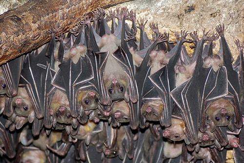
  •  A host species for the Reston strain of Ebola virus that infected and killed a number of laboratory monkeys in Reston near Washington, DC and pigs in Asia has been putatively identified as Geoffroy's Rousette - a fruit bat found across South-East Asia and specifically the Philippines. Provides further evidence of an association of bats and the different Ebola virus species. Read the paper here.
Geoffroy's Rousette fruit bat found across South-East Asia - host of Reston ebola virus?

    Viruses hitch-hike through your body along the immune cell highway

    Nipah virus. 
    Imagine this: Rhinoviruses - one of the culprits responsible for the common cold - enter our body through the upper respiratory tract yet here it stays; the initial and generally the only site of replication is the nasal epithelium. This is how we get a runny/stuffed nose. Contrast this with a virus like nipah virus - a deadly and re-emerging pathogen spread by bats and found across South-east Asia that also enters our body via the upper respiratory tract yet leads to infection and disease in a number of our organs, including the kidneys, blood vessels and the brain, resulting in fatal encephalitis.

    How come one virus remains localised while the other goes systemic? And, more specifically how does it transport itself throughout the body?


    Well, virus infection of a host is a complex multi-step process involving initial contact and entry into the organism (through the nose) , early replication in particular easy-to-access tissues (upper respiratory tract) and then in some cases the spread to specific tissue sites throughout the body (brain). It is these two stages of replication that most often or not lead to the development of disease yet just how does the virus traverse the gap between the two tissue sites - especially given the minute size of a virus particle?

    There are probably three hypotheses of virus spread that could be correct here: lots and lots of virus particles could be released directly into circulation (lymph fluid and blood) from the early site of replication and our blood circulation could do the work for it; the virus could infect those immune cells that cluster around sites of virus infection - or are present naturally in the initial site; or finally, the virus could basically cling on to those highly motile immune cells and be trafficked around the body and effectively transfer infection to the blood vessels and other organs.

    Could your white blood cells transport virus throughout your body?

    One group has recently asked this question with reference to nipah virus (read the paper here) and has discovered that this virus doesn't infect human immune cells although it does bind to them and this virus/cell  interaction facilitates infection of other cells and may allow systemic spread.

    They initially came at this problem at an in vitro level - albeit using cells taken directly from the blood of healthy volunteers. The group must have initially thought that nipah must directly infect white blood cells and this is how it spread - after all this is fairly common for other related viruses, such as measles. To determine exactly which cells supported virus replication they added a green fluorescent protein (GFP) - expressing virus to a panel of white blood cells derived from the blood of the healthy humans and specifically assayed for virus-mRNA synthesis, GFP expression and how much virus was released into the culture medium. Surprisingly only one cell type - dendritic cells (DC's) - an antigen-presenting cell - supported  any kind of replication and even then it wasn't great (see below).

    GFP-nipah virus infects control neuronal U373 cells but not human immune cells - except DC's to an extent

    So how come nipah isn't so good at infecting these cells? Is it a receptor issue? Well the group looked at the mRNA levels of the two nipah virus receptors (Ephrin B2 and B3) in all the cells under investigation and found little correlation between their expression and the ability of nipah to infect them. For example, even the dendritic cell which had the lowest level of both receptors is able to support entry and replication while the other cells (macrophages and monocytes) that express higher levels of it fail to do so. The authors hypothesize that the DC's are engulfing nipah virus particles via a process known as macropinocytosis instead of via virus/receptor binding.

    I mentioned earlier that the virus doesn't actually need to infect the cells to use them as an effective means of transportation - it can really get by through binding to the outer membrane of the cell much like a microbial hitch-hiker. So they looked at how much nipah virus was associated with each cell following stringent washes and surprisingly all the cells looked at were able to bind nipah virus particles even when they failed to get infected themselves. 

    But what exactly is blocking infection when the cells bind virus AND express receptor molecules on their surface - something is inhibiting entry. The paper doesn't really address this issue but points to a role of a virus receptor-independent molecule that binds to nipah virus particles yet prevents internalization and engulfment. And even more interestingly, these virus-laden cells were able to efficiently transfer the infectious particles to other cells - as shown with the DC's and PBL's below and this ability to 'trans-infect' was retained over a couple of days (see below).
    Transfer of infection with virus bound to immune cells overlaid  on top of other cells
    OK, so all this work really paints a nice picture of virus infection in the host through the interactions with certain white blood cells that stably bind to - yet fail to get infected themselves - and hence are able to transfer these infectious particles to other cells throughout the body. But this is all cell culture work - no animals have been worked on here so how are we to know if this actually occurs during infection in vivo? Well, the group performed an experiment where-by the mixed nipah with hamster white blood cells and then injected these virus/cells back in to the animals and finally observed whether disease occurred and if so, how bad was it?

    Hamster infections with nipah or nipah bound immune cells.
    As you can see opposite, by just re-introducing the white blood cells into the animals no death occurred while directly injecting virus into them resulted in 100% mortality but then when the virus/cell mixture was added, these cells were able to transfer the infection to the hamsters with 50% mortality following acute neurological disease reminiscent of that which follows human infection.
    So these cell-bound viruses are able to transmit infection in vivo, which points to it having a role in humans.


    Now with all this information to hand we can now develop a model as to how nipah infects and causes disease following spread within our bodies - this is known as pathogenesis (see below). Virus could initially enter our body through dendritic cells found within certain epithelium (as the virus could infect these cells in vitro) and then these DC's could traffick to local lymph nodes where virus particles could be released and bind to the white blood cells found there; these cells would then go about their business moving around the body thus transferring infection to a range of different cell types (endothelial cells within blood vessels for example)

    Current model for nipah infection and spread within the body

    Just one final thought: watch this video below first - imagine this cell going about its normal routine of moving along your blood vessels and squeezing through them but with it covered in infectious virus; every cell ut encounteers will more than likely be exposed to virus and potentially become infected. No wonder nipah is such a deadly pathogen.


    ResearchBlogging.orgMathieu, C., Pohl, C., Szecsi, J., Trajkovic-Bodennec, S., Devergnas, S., Raoul, H., Cosset, F., Gerlier, D., Wild, T., & Horvat, B. (2011). Nipah Virus Uses Leukocytes for Efficient Dissemination within a Host Journal of Virology, 85 (15), 7863-7871 DOI: 10.1128/JVI.00549-11