Field of Science

Is there such a thing as a completely broad-spectrum antiviral?

I'm sure everyone is aware of the kind of effects virus infection and replication has on the health of humans and other animals (just scroll along my last blog posts and you'll see). 

It's really not good.

And, in most cases we don't have much to prevent or cure it: maybe a vaccine here, some antivirals there yet what we really would like is something that would act against ALL kinds of viruses, from influenzas to smallpox to ebola and even HIV. Most vaccines and antivirals target a very limited number of pathogens, usually only one. The search for this class of antiviral drugs has eluded us in the past yet a paper published this week in PLoS ONE reports (read paper here) the identification of an effective strategy termed Double-stranded RNA (dsRNA) Activated Caspase Oligomerizer or DRACO. See this MIT group's website here.

dsRNA - an important hallmark of viral infection
This approach takes advantage of chiefly two natural processes that occur within cells: the intracellular sensing of long double-stranded RNA molecules and the initiation of programmed cell death, known as apoptosis via what are called as caspase enzymes. Through linking these two pathways together via the construction of a novel protein molecule, this group aim to inhibit viral replication - and hence act as a broad-range antiviral as most known viruses generate dsRNAs.

How and why do cells 'see' ds-RNA?


ds-RNA binding domain from PKR protein
Our cells - and those of nearly every other organism - have a range of defenses to protect themselves from viral infection. One being, the ability to detect when they have been initially infected. For example: through the binding of double- stranded RNA molecules, which viruses produce a lot of. Think of it as a hallmark of virus replication and our cells naturally come equipped with a range of these detector modules that carry out this function using a double-stranded RNA binding motif found somewhere in their structure. Detection of ds-RNA sets off a serious of downstream reactions leading to the cell's normal antiviral response.


Examples:


What about apoptosis?

Apoptosis is a highly coordinated process leading to the self-destruction of the cell without any of the nasty inflammatory responses. The main players in this cellular suicide are the caspases - protease enzymes that specifically target and degrade hundreds of other proteins inside the cell in a well-defined cascade of activity. These proteins are activated through the construction of a larger complexes which themselves are produced as a response to certain noxious stimuli, one example is virus infection. One domain are the death effector domains (an example is a FADD domain) that recruit the initiator caspases into the death-induced signalling complex (DISC) thereby kick-starting the whole process of apoptosis and the cells' death.

The FADD death effector domain for example was added to the end of the ds-RNA binding protein

I said above that virus infection itself activates apoptosis so why doesn't every virus-infected cell just die? Well, viruses have evolved neat tricks to circumvent this response as in most circumstances the death of the cell leads to the end of the infection. The hypothesis that this group are working under is that what if we could couple these two processes more-so than they are naturally as to sidestep the viruses' strategies at combating apoptosis, in a way, short-circuiting this whole pathway. Instead of going through long networks of protein-protein interactions, make it rely only on a single binding. 

So what did they do?

These DRACO proteins are chimeras of the two classes of molecules described above: one half ds-RNA binding and the other a death effector domain. And, finally, in order to get this protein into the cell, they added a 'transduction tag' - a short peptide sequence that is naturally taken up inside cells. So, does it work?

They initially made a whole range of these proteins, using different RNA binding motifs combined with a number of dead effector domains (see below). These recombinant proteins were then tested to see which was the best at getting into cells, binding ds-RNA, triggering apoptosis and most importantly, which was the safest.

The chimeric DRACO proteins (transduction tag in yellow, ds-RNA binding in red and apoptosis in green and blue)

Then they show that these proteins can indeed get into cells and in the presence of ds-RNA they can initiate apoptosis - see below. Also, these proteins were generally well tolerated by these particular cells used.


But does this actually inhibit virus replication?

To test this, they first added their proteins to the cells and then infected them with a virus - in this case human rhinovirus 1B (an important human pathogen) - see data below. Virus infection without administration of DRACO's resulted in 0% cell viability, that is all the cells were killed by the virus. When the proteins were added, viability increased to about 80%. The authors suggest that this because the proteins kill off any virus-infected cells very early thus preventing infection from spreading any further. They also test how effective the strategy is when we add the proteins at different times. It works best when added before infection and then decreases substantially (when added 1 day after infection, viability dropped to 50%).

The antiviral effectiveness of 5 different DRACO proteins in an in vitro rhinovirus infection. The higher the cell viability the better the approach.
Nicely, they extended this same analysis to a very wide range of viruses: dengue, influenza, arenaviruses, adenoviruses, reoviruses - anything they could their hands on. And they all worked much the same as the rhinovirus example above. These viruses are incredibly different from each other, showing just how broadly active this approach is. Different cell lines were also investigated opening the possibility of this approach in a veterinary perspective.

We know that sometimes what works in cell culture doesn't necessarily work in an animal, so the group tried their system in a mouse model of lethal influenza virus infection. When administered prior to challenge, the DRACOs had great results 0 at most 80% of the mice survived, compared to 0 without the proteins. They didn't check out what would happen if they added the DRACOs after infection, which would more closely mimic the situation in real-life.


So, is there such a thing as a completely broad-spectrum antiviral? If there wasn't one before this, there certainly is now after these promising initial results. Although further work will have to establish the exact mechanism of this approach and also apply it to other viruses in more model systems.

To sum up with what the author's say:

DRACOs should be effective against numerous clinical and NIAID priority viruses, due to the broad-spectrum sensitivity of the dsRNA detection domain, the potent activity of the apoptosis induction domain, and the novel direct linkage between the two which viruses have never encountered. We have demonstrated that DRACOs are effective against viruses with DNA, dsRNA, positive-sense ssRNA, and negative-sense ssRNA genomes; enveloped and non-enveloped viruses; viruses that replicate in the cytoplasm and viruses that replicate in the nucleus; human, bat, and rodent viruses; and viruses that use a variety of cellular receptors

ResearchBlogging.org 
Rider, T., Zook, C., Boettcher, T., Wick, S., Pancoast, J., & Zusman, B. (2011). Broad-Spectrum Antiviral Therapeutics PLoS ONE, 6 (7) DOI: 10.1371/journal.pone.0022572

What do the viruses have to say this week? 24/07/11

CDV in macaques
  • A molecular mechanism for the apparently clinically recognised vitamin A-induced inhibition of measles virus is uncovered in vitro. Both Vitamin A and viral infection of cells is required to mediate the inhibitory response (here). Further stresses the importance of preventing vitamin A deficiencies across the developing world. See a major Cochrane review of all measles/vitamin A literature detailing only a significant effect of vitamin A 'megadoses' on measles treatment.
  • A large (>10,000 animals) canine distemper virus (CDV) outbreak in a Rhesus macaque breeding centre in China caused by a unique strain of the virus - reasons how this occurred are unknown but has been going on since 2006. Interestingly, the infection resembled a human measles infection. Paper here.
  • A study investigating the interactions between Herpes simplex virus-1 (HSV-1) - the causative agent of cold-sores - and the nasal mucosal surface was performed using cultured nasal epthithelium taken directly from human patients (read here). The group bbserved viral infection of epithelial cells followed by penetration through the basement membrane and replication in the underlying tissue, shedding light on the mechanisms this virus and other related pathogens use to infect humans and other animals.
Herpes simplex virus -1 spread in human nasal mucosal tissue
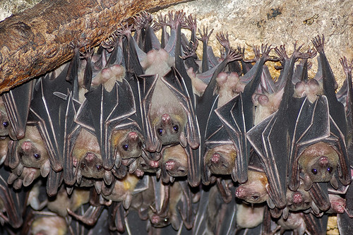
  •  A host species for the Reston strain of Ebola virus that infected and killed a number of laboratory monkeys in Reston near Washington, DC and pigs in Asia has been putatively identified as Geoffroy's Rousette - a fruit bat found across South-East Asia and specifically the Philippines. Provides further evidence of an association of bats and the different Ebola virus species. Read the paper here.
Geoffroy's Rousette fruit bat found across South-East Asia - host of Reston ebola virus?

    Viruses hitch-hike through your body along the immune cell highway

    Nipah virus. 
    Imagine this: Rhinoviruses - one of the culprits responsible for the common cold - enter our body through the upper respiratory tract yet here it stays; the initial and generally the only site of replication is the nasal epithelium. This is how we get a runny/stuffed nose. Contrast this with a virus like nipah virus - a deadly and re-emerging pathogen spread by bats and found across South-east Asia that also enters our body via the upper respiratory tract yet leads to infection and disease in a number of our organs, including the kidneys, blood vessels and the brain, resulting in fatal encephalitis.

    How come one virus remains localised while the other goes systemic? And, more specifically how does it transport itself throughout the body?


    Well, virus infection of a host is a complex multi-step process involving initial contact and entry into the organism (through the nose) , early replication in particular easy-to-access tissues (upper respiratory tract) and then in some cases the spread to specific tissue sites throughout the body (brain). It is these two stages of replication that most often or not lead to the development of disease yet just how does the virus traverse the gap between the two tissue sites - especially given the minute size of a virus particle?

    There are probably three hypotheses of virus spread that could be correct here: lots and lots of virus particles could be released directly into circulation (lymph fluid and blood) from the early site of replication and our blood circulation could do the work for it; the virus could infect those immune cells that cluster around sites of virus infection - or are present naturally in the initial site; or finally, the virus could basically cling on to those highly motile immune cells and be trafficked around the body and effectively transfer infection to the blood vessels and other organs.

    Could your white blood cells transport virus throughout your body?

    One group has recently asked this question with reference to nipah virus (read the paper here) and has discovered that this virus doesn't infect human immune cells although it does bind to them and this virus/cell  interaction facilitates infection of other cells and may allow systemic spread.

    They initially came at this problem at an in vitro level - albeit using cells taken directly from the blood of healthy volunteers. The group must have initially thought that nipah must directly infect white blood cells and this is how it spread - after all this is fairly common for other related viruses, such as measles. To determine exactly which cells supported virus replication they added a green fluorescent protein (GFP) - expressing virus to a panel of white blood cells derived from the blood of the healthy humans and specifically assayed for virus-mRNA synthesis, GFP expression and how much virus was released into the culture medium. Surprisingly only one cell type - dendritic cells (DC's) - an antigen-presenting cell - supported  any kind of replication and even then it wasn't great (see below).

    GFP-nipah virus infects control neuronal U373 cells but not human immune cells - except DC's to an extent

    So how come nipah isn't so good at infecting these cells? Is it a receptor issue? Well the group looked at the mRNA levels of the two nipah virus receptors (Ephrin B2 and B3) in all the cells under investigation and found little correlation between their expression and the ability of nipah to infect them. For example, even the dendritic cell which had the lowest level of both receptors is able to support entry and replication while the other cells (macrophages and monocytes) that express higher levels of it fail to do so. The authors hypothesize that the DC's are engulfing nipah virus particles via a process known as macropinocytosis instead of via virus/receptor binding.

    I mentioned earlier that the virus doesn't actually need to infect the cells to use them as an effective means of transportation - it can really get by through binding to the outer membrane of the cell much like a microbial hitch-hiker. So they looked at how much nipah virus was associated with each cell following stringent washes and surprisingly all the cells looked at were able to bind nipah virus particles even when they failed to get infected themselves. 

    But what exactly is blocking infection when the cells bind virus AND express receptor molecules on their surface - something is inhibiting entry. The paper doesn't really address this issue but points to a role of a virus receptor-independent molecule that binds to nipah virus particles yet prevents internalization and engulfment. And even more interestingly, these virus-laden cells were able to efficiently transfer the infectious particles to other cells - as shown with the DC's and PBL's below and this ability to 'trans-infect' was retained over a couple of days (see below).
    Transfer of infection with virus bound to immune cells overlaid  on top of other cells
    OK, so all this work really paints a nice picture of virus infection in the host through the interactions with certain white blood cells that stably bind to - yet fail to get infected themselves - and hence are able to transfer these infectious particles to other cells throughout the body. But this is all cell culture work - no animals have been worked on here so how are we to know if this actually occurs during infection in vivo? Well, the group performed an experiment where-by the mixed nipah with hamster white blood cells and then injected these virus/cells back in to the animals and finally observed whether disease occurred and if so, how bad was it?

    Hamster infections with nipah or nipah bound immune cells.
    As you can see opposite, by just re-introducing the white blood cells into the animals no death occurred while directly injecting virus into them resulted in 100% mortality but then when the virus/cell mixture was added, these cells were able to transfer the infection to the hamsters with 50% mortality following acute neurological disease reminiscent of that which follows human infection.
    So these cell-bound viruses are able to transmit infection in vivo, which points to it having a role in humans.


    Now with all this information to hand we can now develop a model as to how nipah infects and causes disease following spread within our bodies - this is known as pathogenesis (see below). Virus could initially enter our body through dendritic cells found within certain epithelium (as the virus could infect these cells in vitro) and then these DC's could traffick to local lymph nodes where virus particles could be released and bind to the white blood cells found there; these cells would then go about their business moving around the body thus transferring infection to a range of different cell types (endothelial cells within blood vessels for example)

    Current model for nipah infection and spread within the body

    Just one final thought: watch this video below first - imagine this cell going about its normal routine of moving along your blood vessels and squeezing through them but with it covered in infectious virus; every cell ut encounteers will more than likely be exposed to virus and potentially become infected. No wonder nipah is such a deadly pathogen.


    ResearchBlogging.orgMathieu, C., Pohl, C., Szecsi, J., Trajkovic-Bodennec, S., Devergnas, S., Raoul, H., Cosset, F., Gerlier, D., Wild, T., & Horvat, B. (2011). Nipah Virus Uses Leukocytes for Efficient Dissemination within a Host Journal of Virology, 85 (15), 7863-7871 DOI: 10.1128/JVI.00549-11

    How do viruses hijack our brains to make us vomit - and can we stop it?

    Human rotavirus particles - http://faculty.riohondo.edu
    We've all experienced it; that is, the awful unwell sickness that overwhelms you when you've picked up a nasty viral infection. Remember the last time you had a cold or the 'flu -  when coughs, headaches, sore muscles, vomiting, diarrhea and general fatigue forced you to remain in bed. Yet, just exactly how does the virus manage to do this to you? Especially when in some cases your sickness allowed the virus to spread from you to an uninfected family member; sometimes there is an evolutionary advantage to making you unwell. A recent paper published in PLoS Pathogens this week (which can be read here) investigates the molecular mechanisms behind why the oft' fatal rotavirus causes those infected to vomit and through doing so sheds light on possible therapeutic strategies. 


    A gut villus . http://rezidentiat.3x.ro
    This research isn't just aimed at answering some obscure academic problem - although it does do it - rotavirus induced gastroenteritis is serious business worldwide, with over half a million deaths each year (and many, many more hospitalised) mostly due to severe dehydration caused by the excessive vomiting and diarrhea. In many countries, the rotavirus vaccines just haven't reached the population at large and the reasons why this virus causes vomiting and diarrhea are poorly understood - hence this paper.

    Now, vomiting - like many physiological processes - is controlled by our brain. Usually vomiting is induced by eating something nasty and is hence a kind of defense mechanism to rid ourselves of any harmful things within the gut. In order to sense if there is anything bad in your intestines, the brain is wired up to it's surface through what is known as the enteric nervous system or ENS. This mesh of neurons is connected to particular endocrine sensor cells (see left), called enterochromaffin cells (EC cells) that line our intestines and respond to the harmful stuff through the release of chemical messengers - like serotonin (5-HT), which in turn activate the ENS and end up inducing vomiting and diarrhea via our brain. But, specifically how does rotavirus fit in here?

     A straightforward hypothesis here would be that following ingestion of rotavirus, it would infect the EC cells lining the gut and somehow alter them to release serotonin and stimulate vomiting/diarrhea. And so, the used a primary EC tumour cell line to show that rotavirus does indeed infect the cells. So, now that the virus infects the cell, is this how it causes induction of the ENS? A rapid induction of calcium release was observed soon after infection as well as a quick secretion of serotonin - which itself is dependent on calcium signalling yet the fact that this release occured so soon after addition of  virus suggested that replication was not responsible for it - maybe something in the virus particle or something that was in the solution with the virus was to blame? Following seperation of virus particles from the culture media used to grow it, the group showed that the virus particles themselves were not behind it. The group tracked this effect down to NSP4, a protein secreted from rotavirus infected cells following infection and would therefore have been carried over with the virus in these experiments. In the end it was this protein itself that was responsible for the induction of calcium signalling and downstream serotonin release. 

    Gut villi from mice infected with rotavirus. Arrows indicate rotavirus infected cells (those with large vacuoles) and stars indicated EC cells stained brown. Below, red = rotavirus and green = serotonin and yellow = colocalisation or a rotavirus infected EC cell. Yellow cells are rare - non-EC infected cells (enterocytes) are not.

    All done and dusted now? No - up to this point, all work was done under tissue culture conditions in vitro - all be it, mostly in primary cells. The group then moved on to a small-animal mouse model of rotavirus infection to determine if this was the case in vivo - does rotavirus infect EC cells and induce calcium/serotonin signalling that stimulates gut neurons to induce vomiting/diarrhea. This could not have been addressed in cell culture. Following mouse infection, cross-sections of the gut wall were studied to determine the location of rotavirus infected cells and EC cells (see above). Many of the enterocyte cells lining the mouse gut were infected while only rarely were the EC cells targeted - despite what was observed in vitro. Yet for this model to work, rotavirus did not require infection of EC cells as NSP4, the protein behind this effect is secreted from cells and could hence stimulate EC from a distance. In the mouse, the group show that serotonin administration results in diarrhea (oddly, mice do not - and cannot - vomit) and also rotavirus infection leads to the activation of regions in the brain associated with sickness in humans (done through fos staining of brain sections).

    A model for how rotavirus makes us vomit/diarrhea
    This work has built up a kind of nice and simple picture of a mechanism of rotavirus causes vomiting and diarrhea. They have shown that the virus is able to enter and replicate inside epithelial cells and endcocrine cells lining the gut; they showed that this led to the release of serotonin - the stimulator of vomiting/diarrhea - and that this was down to NSP4 synthesis and release and finally that mice infected with rotavirus activated brain regions associated with vomiting and diarrhea.

    The model: (see above) - Rotavirus infects gut cells - expresses and releases NSP4 - NSP4 stimulates calcium signalling and serotonin release from nearby EC cells - serotonin stimulates close by neurons that activate vomiting/diarrhea areas of the brain, which causes serious fluid loss, dehydration and subsequent death. Also this results in rapid spread of virus particles and transmission of infection.

    The good thing is now, with this mechanism at hand, we may be able to partially inhibit this response and save the lives of many children and prevent the spread of rotavirus in these populations. Luckily for us, a number of generic - and hence cheap - already in use anti-sickness tablets (serotonin receptor antagonists) exist and could be applied in this setting. Although previous research had established the efficacy of using this drug to attenuate diarrhea in rotavirus infections, this paper established a working mechanism for its activity.

    Granisetron - a serotonin receptor antogonist used as an anti-sickness medication and could be developed to aid rotavirus-induced vomiting/diarrhea. http://upload.wikimedia.org/wikipedia/


    ResearchBlogging.orgHagbom, M., Istrate, C., Engblom, D., Karlsson, T., Rodriguez-Diaz, J., Buesa, J., Taylor, J., Loitto, V., Magnusson, K., Ahlman, H., Lundgren, O., & Svensson, L. (2011). Rotavirus Stimulates Release of Serotonin (5-HT) from Human Enterochromaffin Cells and Activates Brain Structures Involved in Nausea and Vomiting PLoS Pathogens, 7 (7) DOI: 10.1371/journal.ppat.1002115

    Vaginal 'probiotics' may protect against HIV transmission

    According to WHO data, 2009 saw 33.9 million people worldwide infected with the human immunodeficiency virus -  HIV and of these, 1.8 million died from AIDS while 2.9 million were newly infected with the virus. This leaves a year-on-year increase of just over 1 million HIV positive people of which, many of these will go on to pass the virus. Therefore any strategy to eliminate HIV from the human population will have to aim at both treating those already infected as well as preventing new viral transmission and if this is achieved, HIV infection worldwide would dramatically decrease with every year, severely reducing the global health burden caused by this viral pandemic. The only problem is, how can we successfully prevent transmission?

    Despite a significant decrease the number of people infected by HIV continues to grow year on year

    HIV entry and pathogenesis - where and how can we target it?

    One of the major routes of HIV transmission is through sexual activity: an HIV positive person will be carrying many copies of the HIV genome within their cells and in turn these will be able to generate new infectious virus particles. These virions - either present in blood or semen - may be mechanically transferred from infected to uninfected people during sexual activity and many immune cells (Langerhans cells, macrophages and intraepithelial CD4+ T cells to be precise - see below) lining and within the mucosal epithelial surfaces of the reproductive tract are the initial target cells for HIV entry into the human body. It is here that the cells are exposed to the virus through sexual contact, facilitating virus uptake and initial infection then allowing the virus to spread within the body and potentially set up the chronic infection that may develop into AIDS.
    HIV entry via immune cells within the vaginal mucosa and spread to systemic lymphoid organs. Miller, 2007.

    Many potential strategies are currently in development which aim at preventing person-person transmission during sexual activity through the inhibition of HIV particle transfer. This is the reason why condoms - male and female - are so effective. As the authors of the paper outlined below highlight the requirements of such a strategy:
    In many cultural settings, women need a product that can be used covertly without obtaining the permission of their sexual partner. In addition, the cost of HIV prevention must be affordable to the developing world. Thus, there is still a need for products that block HIV transmission, are safe and easy to use, and are coitally independent, discreet, and cost effective.
    Bacterial symbiosis to the help

    All being so, our bodies are not completely defenseless when it comes to preventing sexually transmitted infections, including HIV; we have multiple tricks up our sleeves and one of which is through a form of bacterial symbiosis. The human reproductive tract is covered in a bacterial biofilm composed of a few species of bacteria, predominantly Lactobacilli. These microbes regulate vaginal biology and aid in the protection against variable infections through the formation of a physical barrier and through alterations in pH. But of course it isn't enough and as I mentioned earlier, people are all too often getting infected with HIV. Yet what if we could enhance these bacterial defenses through genetically engineering those bacteria that natural colonise the reproductive tract? This is exactly what a recently published paper in Mucosal Immunology reports -  the development of a novel genetically engineered live bacterial strain expressing an anti-HIV protein that can be easily applied to the vagina and prevents HIV transmission

    Normal tissue properties A) and Presence of anti-HIV protein CV-N

    The group had previously generated an engineered strain of Lactobacillus jensenii (termed:1153-1666) that expressed and secreted a modified Cyanovirin-N (CV-N) protein. This protein has been shown to have a broad inhibitory activity against a range of HIV-1 strains and provide protection from infection in a non-human primate model of HIV through inhibiting virus entry into those target immune cells - it is also highly potent and non-toxic. To establish the potential for administering this bacteria in humans, the group inoculated the vaginas of non-human primate macaques on a regular basis and achieved stable colonisation along with expression of the antiviral protein in the fluid lining the tract; the recombinant L. jensenii was detected along the epithelium while no tissue changes were observed and no untoward inflammation was detected, all indicating the safety of this strategy.

    in vitro HIV inhibition with non-recombinant (left) and recombinant (right) bacteria

    So far, the ability of this recombinant bacteria to inhibit HIV infection has not been addressed so later it was assessed through the use of an in vitro model that accurately portrays the physical and biological architecture of the human vagina as well as the macaque model that uses chimeric human/simian immunodeficiency virus infection. Colonization of the in vitro vaginal tract with the non-recombinant bacteria resulted in a 23% reduction in HIV infection compared to the control while inoculation with the CV-N expressing strain gave 72% inhibition (see above). Groups of macaques were then treated with an antibiotic to remove any endogenous Lactobacillus colonizing their vaginal tract and were subsequently inoculated repeatedly with the recombinant strain. These monkeys were then repeatedly challenged with the virus in a manner similiar to what would happen under natural human conditions and the ability of the virus to infect each animal was assessed over time. This strategy reduced the infection rate by nearly 63% (see below).

    Macaque challenge outline and results of bacterial colonization protection


     This group has generated a bacterial strain - closely related to that already present within the human reproductive tract - that has been genetically augmented through the introduction of the gene for a potent HIV-inhibitory protein Cyanovirin-N. This recombinant bacteria can be inoculated into the vagina and affords protection against initial HIV infection in an in vitro and in vivo macaque model through the expression and secretion of CV-N without generating a toxic response; this engineered microbe is thus better at preventing infection that the non-CV-N expressing bacteria. It remains stable over time But, how then would this function in the real world?

      In order to halt the HIV pandemic, we require an at least partially effective strategy to prevent person-person transmission of the virus and this method must safe and inexpensive if it is to be administered to the many people who require it across the developing world. This method outlined above - pending the results of further clinical work - highlights the importance of novel strategies to combat HIV spread. The group outlines the development of an easy-to-apply, safe, efficacious and cheap method to combat HIV transmission in two model systems. Despite these pleasing results, further work will need to be carried out in a clinical field trial to determine whether this mode of protection will work in the real world.


    ResearchBlogging.orgLagenaur, L., Sanders-Beer, B., Brichacek, B., Pal, R., Liu, X., Liu, Y., Yu, R., Venzon, D., Lee, P., & Hamer, D. (2011). Prevention of vaginal SHIV transmission in macaques by a live recombinant Lactobacillus Mucosal Immunology DOI: 10.1038/mi.2011.30

    Part II - why vaccine viruses are weaker that their pathogenic cousins

    I've talked about this before (here with mumps virus) but just what makes vaccines so good at being vaccines? - that is, what makes them safe yet immunogenic? The great thing about this is that maybe if we understand these processes a bit better we may even be able to develop safer, more effective and cheaper vaccines that will ultimately save more and more peoples lives. A lot of research at the minute is cuurently attempting to tease apart the roles of hundreds of virus genes in infection and disease with many groups focusing on specifically how these genes affect attenuation, that is how they make the virus weaker and less dangerous so it can be used as a vaccine.

    So it starts off like this: we know vaccines are safe and effective when after we give them to people/other animals very little get sick and a lot of them are immunized against infection with that particular virus yet the 'wild-type', normal virus is able to infect and cause disease - so just what makes these different? Well to find out we have to compare the two viruses - the weak one and the virulent one and somewhere in their genomes lie differences that will possibly shed light on the important molecular mechanisms behind virus attenuation and pathogenesis.

    Canine distemper in dogs. http://www.dogs-info.net
    In a recent paper, Dietzel et al investigated these processes using a great model system, specifically canine distemper virus - or CDV, a virus able to infect and cause life-threatening disease in a whole range of mammalian species in a domestic or wild-life situation and hence is obligatorily given as part of a multivalent vaccine to all domestic dogs. The group began by comparing the matrix - or M- gene between a vaccine strain of a virus known as canine distemper virus or CDV and a wild-type one. This M gene encodes a protein which plays a major role in the assembly of new virus particles, cell-cell spread and particle stability within CDV-infected cells through co-ordinating genome interactions with virion-surface proteins. Its role in CDV pathogenesis - and other viruses - has barely been looked at. The two M proteins differed at only six different amino acid positions (3%) so to look into the role of them both during CDV infection the group genetically engineered a CDV virus based on the wild-type but carrying the vaccine-derived M gene in place of its own. The growth and physical characteristics of the three viruses :wild-type, wild-type + vaccine M and vaccine strain viruses were looked at as well as physically characteristics of the produced particles, how they are assembled and what happens during animal infection.
    
    The 3 viruses grow equally well in cell culture conditions.
    
    Despite the three viruses growing equally well under cell culture conditions (see growth above and how the infection looks in tissue culture - all are pretty much similiar), the two differed markedly in their particle-infectivity ratio, that is - the number of virus particles capable to carry out a successful infection compared to the total number of particles in a given volume, i.e some particles may be non-functional for some reason, maybe because they are less physically stable or damaged.

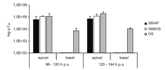
    virus particle release from the tops or bottoms of the cells

    The vaccine virus also differed in the way new CDV particles were released from cells: the wild-type virus is released from the tops of polarised epithelial cells while the vaccine strain is released from both the top and the bottom. However, transfer of the vaccine M gene wasn't able to confer bipolar release onto the wild-type virus indicating the function of other CDV genes in this phenotype. To determine a mechanism for this bipolar release the group analysed the intracellular distribution of the key CDV proteins, H and F involved in particle production and cell-cell spread. They found that this proteins differed in their location between the three viruses: the wild-type only had them on the tops of cells while the vaccine strain had them on both sides, the chimeric virus had an intermediate distribution. These results highlight the primary ability of the CDV matrix protein to coordinate protein distribution within the cell.

    CDV protein intracellular distibution


    All these results are good and all but what effect do they actually have in vivo - in a situation where the virus is infecting multiple cell types and is constantly battling the immune system? To determine the effect of vaccine M gene, groups of ferrets (an excellent model of CDV infection that are naturally infected by the virus) were inoculated intranasally with the three viruses and disease severity, body temperature, disease symptoms and fatality was recorded. Interestingly, based on these measures, the wild-type with the vaccine M was more attenuated that the vaccine virus as shown below with the measure of ferret blood leukocytes - the more blood cells the less pathogenic the virus.

    A measure of CDV disease - the percent of ferret blood cells - a CDV target cell
    
    The process of pathogen attenuation for the generation of protective vaccines has worked well in the past for many diseases yet many viruses and bacteria do not prove to be responsive to this method. Other more targeted processes may have to be designed in order to create vaccines for against these diseases. Work investigating the molecular basis of virus pathogenesis and attenuation may allow us to rapidly and successfully attenuate these hard-to-vaccinate viruses in a more targeted fashion - and one that may work better than the previously tried methods.

    ResearchBlogging.orgDietzel, E., Anderson, D., Castan, A., von Messling, V., & Maisner, A. (2011). Canine Distemper Virus Matrix Protein Influences Particle Infectivity, Particle C
    omposition, and Envelope Distribution in Polarized Epithelial Cells and Modulates Virulence Journal of Virology, 85 (14), 7162-7168 DOI: 10.1128/JVI.00051-11


    6cfa3635b7da201097d1cb415b03572d

    How come vaccine viruses are so safe while normal viruses are so dangerous?

    One method that has been used extensively to generate worthwhile vaccines is that of forcing an initially disease-causing virus to replicate inside a non-natural cell, for example imagine forcing the human specific measles virus to replicate within cells from a chicken. Over time these viruses - all with extremely high mutation rates - will evolve and adapt to the conditions within a chicken cell while at the same time losing it's ability to survive within human cells. The use of these live-attenuated viruses as vaccines has led to the dramatic reduction in a number of important human - and livestock - viruses - the likes of measles, mumps, polio and rinderpest. 

    These vaccine viruses usually retain their ability to infect and replicate within their host (one of the reasons why they are so good at protecting us) yet fail to cause significant disease past the odd fever. However, in some cases the use of these vaccines has led to a number of cases where they caused serious illness. Hence, with the powerful immunity these vaccines generate comes the important potential chance of causing disease and therefore the ability to understand and predict how a particular vaccine will behave following administration is key to continuing the safe use of live attenuated vaccines. Sadly, our knowledge of the mechanisms behind virulence and attenuation are largely unknown.

    Vaccination of the man behind the mumps vaccine - Maurice Hilleman's daughter, Kirsten, while her half-sister, Jeryl Lynn, looks on. The MuV vaccine strain Jeryl-Lynn was originally developed from the virus which infected Hilleman's daughter of the same name through non-natural replication. http://www.historyofvaccines.org/

    One example of this can be found with the case of the mumps virus (MuV) vaccine. Interested in mumps? see here. Before the widespread use of the vaccine, MuV was responsible for the majority of cases of aseptic meningitis (inflammation of the lining of the brain not caused by bacteria) in the western world - this virus is highly adapted in entering the human central nervous system and replicating within the epithelial cells that line its inner layer, making it particularly dangerous. Because of this, any vaccine batch produced must go through rigorous pre-clinical testing, the majority of which is carried out in primates. Yet sometimes, vaccine viruses will lead to the development of aseptic meningitis following immunization. We still do not adequately understand the molecular basis of the ability of MuV to cause disease in the nervous system - something that may facilitate the development of new and improved MuV vaccines.

    Sauder et al, publishing in Journal of Virology, explore the genetic basis of MuV neuropathogenesis through the generation of around 30 chimeric viruses - see below - comprising genes of a neurovirulent MuV (strain: 88-1961) and a highly attenuated MuV (vaccine strain Jeryl-Lynn 5). Through the analysis of the potential for these viruses to cause disease in a rat model of mumps meningitis they were able to assess the contribution of specific genes - or combinations of genes - in either increasing or decreasing its ability to cause disease.

    Chimeric mumps viruses (combinations of attenuated - JL and virulent - 88). What effect will each have on pathogenesis?

    MuV has a single-stranded RNA genome of 15,384 nucleotides and encoded within this one molecule are 7 genes through which at least 9 different proteins are expressed.  These proteins - and hence their corresponding genes - govern the basic biology of this virus: building of the virus particle, receptor binding, cell entry, transcription and replication and finally, cell exit. N, P and L forming the replication apparatus of the virus while M, F, SH and HN are involved in particle assembly, entry and exit. It is these same proteins that are responsible for the ability of MuV to cause disease in humans, specifically aseptic meningitis. Any understanding of the ability of a virus to cause disease must identify its key molecular - and genetic - components hence, what particular genes along the MuV genome are responsible for causing aseptic meningitis in humans? Is it those that allow the virus to enter the cell? Those involved in replication? or those involved in building the virus particle? Sauder et al sought to try and convert an attenuated virus into a virulent one and vice versa - and in doing so uncover the biology behind MuV pathogenesis.
    Can we transform the virulent 88 strain to an attenuated virus by inserting combinations of attenuated JL genes?


    Above shows the results from the initial experiment comparing disease caused by the different viruses: This is where the added different genes from the attenuated virus to the virulent to see whether or not this weakened the viruses ability to cause disease. As you can see, no individual genes or combinations added to the 88-1961 virus caused it to be as attenuated as the vaccine strain, suggesting that in this case with MuV attenuation is a complex, polygenic trait involving many genes. Neither the transfer of all replication proteins (N, P and L) nor the assembly proteins (M, F, SH and HN) resulted in complete attenuation. The most dramatic effect was seen with N and M transfers - although the reason why was not addressed. 


    Can we transform the attenuated JL strain to a virulent virus by inserting combinations of pathogenic 88 genes.

    Following on from this, the group tried to see whether they could turn the attenuated into the virulent virus through carrying out the reverse of the above experiment although this time the results were not the same as no genes or combinations resulted in anywhere near the levels of pathogenesis seen for the 88-1961 virus. Even with the addition of the previously effective N and M combination.
     
    These results tell us a number of things about MuV attenuation and virulence, firstly: it's a lot more complex than we might have previously thought! - this is not all down to one gene but a few of them working together in combination. Secondly, it doesn't work both ways - the mechanisms behind how a virus causes and doesn't cause a disease are different as the same genes couldnt do both. And lastly these results point us to some interesting avenues of future research into virus attenuation - what are the specific molecular roles each of these genes play in turning the virulent virus into an attenuated one? For example, why do the attenuated N and M have such a drastic effect? More work will be undoubtedly be done to determine these questions and this may allow us to rationally attenuate viruses in the future using these combinations of genes.

    ResearchBlogging.orgSauder CJ, Zhang CX, Ngo L, Werner K, Lemon K, Duprex WP, Malik T, Carbone K, & Rubin SA (2011). Gene-specific contributions to mumps virus neurovirulence and neuroattenuation. Journal of virology, 85 (14), 7059-69 PMID: 21543475

    Can we use viruses to vaccinate against - and cure - established cancers?

    Everyone is aware of the ability of our immune system to defend against microbial pathogens yet its role in the prevention of other diseases - like cancer - is generally over-looked. Yet it is through the harnessing of our immune system that novel ways of combating cancer may arise. And interestingly enough, through the use of engineered viruses - the same ones our immune system protects against - we may now control aspects of immunity to suit these medical needs. Kotke et al, report in Nature Medicine just this week, their use of a new virus-based immunotherapy platform that was able to effectively 'cure' mice suffering with cancer.

    One of the hallmarks of cancer appears to be the ability to persist in the face of an active immune system - see fig. 1. Newly cancerous cells and tumours are able to survive and proliferate without - or at least protecting themselves - against a full-blown immune attack. Our immune system is usually able to protect us from the development of cancer but in some cases something fails and the result is more often than not - cancer.

    Fig 1. Newly recognised cancer hallmarks - note avoiding immune destruction. (Hanahan and Weinberg 2011).


    Through the recognition of 'tumour antigens' (proteins expressed only on cancer cells) or 'tumour-associated antigens' (proteins expressed differently on cancer cells), our immune system is usually able to mount an effective response toward those cells. This is the  system that tumours are able to suppress yet we may be able to boost the natural immunological nature of tumours in order to cure them. The discovery of these proteins - just like those found on the surface of virus particles or bacterial cells - may allow us to effectively vaccinate people against cancer, allowing their own immune system to remove the cancerous cells.

    This is what we try and achieve through cancer vaccines and immunotherapies. Kotke et al set about trying to improve upon these current immunotherapy platforms, which - as they state - suffered from a number of problems including: lack of known tumour antigens and coverage of only a few such proteins. Most current immunotherapies rely upon the immunization with only a single antigen. Previous work by the group showed that if you kill normal cells from a patient in vivo - you may be able to elicit an effective anti-tumour immune response through the induction of tumour-associated antigen immunity (when cells die they either burst and release their insides).This work effectively showed that you could immunize with a wide range of tumour-associated antigens from normal cells and protect against cancer - both circumventing the above two problems.


    VSV particles - www.standford.edu
    To improve upon this model, they developed a virus-based platform for the expression of a wide range of tumour-associated antigens in vivo termed altered self antigen and epitope library (ASEL) - see fig 3. They based their method upon the vesicular stromatitis virus - or VSV - normally a virus solely of livestock that also has the ability to infect humans. In humans it causes a generally mild flu-like illness and may form vesicles on the skin. Although a single-stranded non-segmented negative sense RNA virus, we have the ability to generate infectious VSV particles entirely from cDNA plasmids encoding the entire VSV genome. This has facilitated the development of VSV as a key eukaryotic expression vector for multiple uses, such as these cancer immunotherapies and specifically, as an oncolytic treatment. Using standard molecular biology techniques (PCR, restriction enzyme digests and ligations) you can insert any gene from whatever source you want into the VSV genome and it will be expressed inside cells following infection. The benefit with using this virus is that even without the expression of tumour antigens from it's genome, replication within a cell will kill the cell anyway. It is a double hit strategy.

    Fig.3. Cloning the cDNA library into the VSV genome in forward and reverse orientations = VSV-ASEL library

    In order to express hundreds of tumour-associated antigen genes, the group used reverse-transcriptase PCR to amplify all expressed genes from normal prostate tissue and inserted the entire normal prostate tissue cDNA library into VSV. They were then able to infect mice that suffered with prostate cancer and observe what happened to their tumours - specifcally, was an effective immune response generated and did the tumour shrink? VSV virus particles were injected into the mice, virus entered the cells of the mice and began to replicate and express their genes, including the newly inserted prostate cDNA. Essentially, thousands of virus particles were adminsired, each containing a slightly different gene from the prostate cDNA library. High levels of tumour associated antigens were therfore being expressed in mice allowing for the generation of an effective immune response.

    Survival of mice treated with the VSV viruses - GFP expressing negative control; and the VSV ASEL in mice with established 'TC2' prostate tumours.

    This approached effectively cured the mice who suffered from prostate cancer. Following this treatment a number of resistant tumours emerged which were again subjected to a further treatment using a cDNA library taken from the tumour itself this time and this readily treated the secondary resistant cancers. The clinical benefits of this approach can hardly go unnoticed. The ability to administer a broad tailored therapy that has the potential to cure an established tumour will be revolutionary, especially given the relative ease at which this can be developed 'at the bedside'. The ability to easily genetically manipulate viruses has - and will continue to - revolutionise the medical sciences. Look out for the eminent clinical trials. 


    ResearchBlogging.orgKottke, T., Errington, F., Pulido, J., Galivo, F., Thompson, J., Wongthida, P., Diaz, R., Chong, H., Ilett, E., Chester, J., Pandha, H., Harrington, K., Selby, P., Melcher, A., & Vile, R. (2011). Broad antigenic coverage induced by vaccination with virus-based cDNA libraries cures established tumors Nature Medicine DOI: 10.1038/nm.2390

    Improving global immunisation with more heat tolerant vaccines

    During the last century, mass vaccination campaigns were rolled out across much of the industrialised world. And, with much success, we have near eradicated most common virus infections from these populations.   Despite the more recent efforts to bring the developing world into this fold, major setbacks have been uncovered, one being: how exactly are we to stably transport  delicate vaccine stocks from their place of production to the regions that need it most.

       Many of these vaccines, known as 'live attenuated vaccines' - or LAVs-are generally unstable in the environment outside of our cells and bodies. For example, the measles virus has an outer lipid membrane that is highly delicate and damage to this removes any chance of this vaccine working. Basically, these LAVs, which rely upon a weaker infection, cannot successfully complete their replication cycle without an envelope. Generally, the ability of a vaccine to provide protection is intrinsically linked to its overall structure; you destroy the structure, you destroy protection.

    Unstable measles virus particles
      The more difficult places to transport vaccines to  happen to be more tropical regions, like sub-Saharan Africa, South America and South-East Asia. These places are very warm and humid which goes against what our vaccines like. With temperatures up to 40 degrees Celsius and the fact that freezing also destroys the vaccine, the only way forward is the implementation of a cold chain. If we keep the vaccine stock 'chilled' from manufacture (see Merck or the Serum Institute of India) all the way to administration, then we have a chance to prevent its degradation. But this is not as easy as it may sound as it is economically and logistically difficult to achieve this in developing countries. To overcome this, the produced vaccine stock is freeze-dried to remove any water that may contribute to its instability and when this prep reaches the clinic it is then reconstituted through the addition of a solvent preparation, which has then all got to be kept cool.


    With measles virus vaccines however, even upon reconstitution, there are still major losses to immunogenicity and this problem is compounded with the use of multi-dose vials of vaccines. A single vaccine stock, prepared in the morning may now sit in the clinic unrefrigerated until the end of clinic hours. If we were then to get immunized at 5:00 pm, what are the chances of that being successful? It is thus not much of a surprise to hear that many virus outbreaks have been attributed to breaks in the cold chain like this. Our ability to eradicate many diseases in these countries and hence worldwide, is blocked by difficulties in the cold chain.

       Being so, Bill and Melinda Gates identified "Vaccines That Do Not Require Refrigeration" as one of their funded 14 Grand Challenges in Global Health and the results of one potential solution to this problem have just been published in the journal Vaccine (read it here). This work aimed at identifying novel reconstitution liquids that would increase the thermostability of the prepared vaccine and therefore increase its 'shelf-life' in these more difficult environments. Using a recombinant measles vaccine virus that expresses green fluorescent protein (GFP) upon infection, the group were able to assess the level of infectivity of a range of vaccine stocks reconstituted in varied solutions.

    Counting measles virus infected (GFP positive) cells


       Indeed, they tested >11,000 formulations (myriad combinations of buffers, stabilizers, solubilizers, preservatives, pH and tonicifiers - whatever they are?) using their recently developed high-throughput system in which they were able to automate the whole process. These results were validated and confirmed with another virus, this time an adenovirus expressing GFP; results were the same. The group identified a formulation that caused the vaccine to "suffer <1.0 log loss after 8 h at 40 ◦ C in the liquid state", a major improvement on a previous loss of" 1 log of potency after 8 h at 37 ◦ C in the reconstituted (liquid) form". In conclusion, they identified a novel vaccine formulation that substantially increased the stability of two viruses, which can be used in immunization campaigns worldwide. As I've alluded to before, the use of high-throughput screens has much potential in the world of virus research.
    Log loss of infectivity of vaccines


       How is this result likely to change vaccine implementation in the real world then? Well, the evidence presented here indicates the ability of creating more stable vaccines that would transfer to better vaccine coverage yet the real problem appears to be whether current vaccine manufacturers would accommodate such a significant change to production, especially given their inherent unwillingness to invest in the developing world.

       The cost associated with this may be offset by the savings made through reduction in vaccine wastage and loss of cold chain implementation but we must realise that only when such an improvement looks attractive to the market will change come. The future of vaccination looks to be bright, especially with the combination of newer technologies and the backing of large philanthropic organisations.


    ResearchBlogging.orgSchlehuber LD, McFadyen IJ, Shu Y, Carignan J, Duprex WP, Forsyth WR, Ho JH, Kitsos CM, Lee GY, Levinson DA, Lucier SC, Moore CB, Nguyen NT, Ramos J, Weinstock BA, Zhang J, Monagle JA, Gardner CR, & Alvarez JC (2011). Towards ambient temperature-stable vaccines: The identification of thermally stabilizing liquid formulations for measles virus using an innovative high-throughput infectivity assay. Vaccine PMID: 21616113